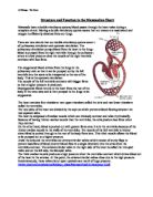
The muscle of the heart is called cardiac muscle and is made of tightly connecting cells. This close contact allows rapid ion transport from cell to cell. This then allows smooth, efficient waves of depolarisation to produce contractions (and repolarisation to bring about relaxation), which pass through the heart. The tissue is said to be myogenic, i.e. it does not need electrical impulses from a nerve to make it contract. If the cardiac muscle is supplied with oxygen and nutrients (a task carried out by the coronary arteries which you can see running over the surface of the heart) it will continue to contract at a steady pace. Nerves supplying the heart, though they are not needed to start the contractions, can bring about an increase or decrease in the rate of contractions when appropriate. The wall of the heart is made up of three layers: the endocardium, which is smooth tissue inside the heart to help blood flow, the myocardium, which is the striped cardiac muscle and strong pumping part of the heart, and finally, the epicardium, which is the outer layer of tissue and links with the pericardium (a double layered bag surrounding the heart).
One cardiac cycle consists of the atria and then the ventricles contracting so that the blood that has entered the heart is pumped out. This occurs about 72 times every minute and is continuous. One heartbeat lasts for about 0.8 seconds. The periods of contraction are called systole and make up about 0.3 seconds; the periods of relaxation are called diastole and last about 0.5 seconds. The basic heart rate can be modified by the nervous system according to the demands of the body. E.g. during heavy exercise, the heart rate and the breathing rate will increase significantly.
When the atria and ventricles are in diastole, blood at a low pressure in the veins flows into the atria which fill with blood and increase this pressure, meanwhile the atrioventricular valves are shut. With rising atrial pressure these valves are then pushed open and the ventricles start to fill with blood. During the 0.3 seconds of contraction, there is both atrial systole followed by ventricular systole. The sinoatrial node initiates an electrical impulse that travels across the atrial walls. This contracts the myocardium of each atrium and atrial systole then forces all remaining blood past the atrioventricular valves into the ventricles. The semi-lunar valves are closed during this. The electrical impulse passes from the atrioventricular node to the bundle of His then to the purkinje fibres. There is a slight delay at the AV node to ensure that both contract at the same time. The myocardium of the ventricles contract, the bicuspid and tricuspid valves remain closed, and the pressure increases in the ventricles and pushes open the semi-lunar valves. This whole contraction results in blood flow out of the heart and into the pulmonary and systemic circulatory systems.
Cardiac contractions are initiated by an electrical impulse that originates from the sinoatrial node (pacemaker). The waves spread out over the two atrial walls so that they contract. There is a band of fibres between the atria and ventricles, which have a high electrical resistance so the waves cannot spread from the atria to the ventricles. The impulse travels to the atrioventricular node situated in the atrial septum. The impulse then spreads into a specialised tissue known as the bundle of His, which connects the AV node to the branch network of purkinje fibres, located in the septum and ventricle walls. The purkinje fibres are connected to cardiac muscle fibres (myocardium) therefore the impulse spreads throughout the ventricle walls causing contraction.
Electrical activity of the heart:
P = Excitation of both the atria – SA node – AV node
QRS = Excitation of both ventricles. AV node – bundle of His – purkinje fibres
T = ventricles as they relax
A person’s heart rate is controlled by three factors: neural control, hormonal control and intrinsic control. The autonomic nervous system and the sympathetic and parasympathetic nerves that stimulate it control the SA node. These nerves are stimulated in various situations, e.g. during exercise, the accelerator nerve is stimulated. It releases noradrenaline at the SA node resulting in the heart rate increasing due to a decreased delay at the AV node and increasing the force of the contractions. One nerve, the accelerator nerve, runs from the cardioacceleratory centre in the medulla of the brain to the SA node. Another, the vagus nerves, runs from the cardioinhibitory centre in the medulla of the brain to the SA node. If the vagus nerve is stimulated, acetylcholine is released at the SA node. The delay at the AV node increases and the cardiac output falls. Blood pressure also affects the cardiac output. Some blood vessels (e.g. the aorta and carotid arteries) have stretch receptors in their walls. These detect the pressure and send impulses to the cardiac centre in the medulla. If the pressure is too high: the cardioinhibitory centre is stimulated, impulses are sent down the vagus nerve, the heart rate is slowed and the pressure will fall. If the pressure is too low: the cardioacceleratory centre is stimulated, impulses are sent down the accelerator nerve, the heart rate is increased and the pressure will rise. Hormones can control heart rate when adrenaline is released from the adrenal medulla, flows in the blood and affects the SA node. The SA node is stimulated, works faster, increasing the heart rate.
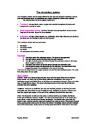
After leaving the heart, deoxygenated blood goes to the lungs to pick up oxygen and oxygenated blood is transported around the rest of the body through the blood vessels. Arteries are blood vessels that carry blood away from heart. Arterial walls are able to expand and contract. Arteries have three layers of thick walls. Smooth muscle fibres contract, another layer of connective tissue is quite elastic, allowing the arteries to carry blood under high pressure. This is the structure of an artery:
The aorta is the main artery leaving the heart. The pulmonary artery is the only artery that carries oxygen-poor blood. The pulmonary artery carries deoxygenated blood to the lungs. In the lungs, gas exchange occurs, carbon dioxide diffuses out, oxygen diffuses in. arterioles are small arteries that connect larger arteries with capillaries. Small arterioles branch into collections of capillaries known as capillary beds.
Capillaries are thin-walled blood vessels in which gas exchange occurs. In the capillary, the wall is only one cell layer thick. Capillaries are concentrated into capillary beds. Some capillaries have small pores between the cells of the capillary wall, allowing materials to flow in and out of capillaries as well as the passage of white blood cells. Nutrients, wastes, and hormones are exchanged across the thin walls of capillaries. Capillaries are microscopic in size, although blushing is one manifestation of blood flow into capillaries. Control of blood flow into capillary beds is done by nerve-controlled sphincters. This is the capillary structure:
The extensive network of capillaries in the human body is estimated at between 50,000 and 60,000 miles long. Blood leaving the capillary beds flows into a progressively larger series of venules that in turn join to form veins. Veins carry blood from capillaries to the heart. With the exception of the pulmonary veins, blood in veins is deoxygenated. The pulmonary veins carry oxygenated blood from lungs back to the heart. Venules are smaller veins that gather blood from capillary beds into veins. Pressure in veins is low, so veins depend on nearby muscular contractions to move blood along. The veins have valves that prevent back-flow of blood. The structure of veins:
As blood gets farther from the heart, the pressure likewise decreases. Each contraction of the ventricles sends pressure through the arteries. Elasticity of lungs helps to keep pulmonary pressures low. Systemic pressure is sensed by receptors in the arteries and atria. Nerve messages from these sensors communicate conditions to the medulla in the brain. Signals from the medulla regulate blood pressure.
In these blood vessels vital substances are transported in the blood to the tissues around the body. This is for processes such as respiration to take place. Substances include glucose, oxygen taken to the tissues, and carbon dioxide and metabolic waste, which are taken back from the tissues as waste products.
Bibliography:
Advanced Biology 1
Biological Sciences 2