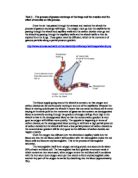
Once the oxygen has diffused over the alveoli and capillary walls into the blood and into the red blood cells it will combine with the haemoglobin inside the red blood cells and become oxyhaemoglobin. This is the process of haemoglobin saturation.
The haemoglobin itself is an oxygen carrying protein and accounts for about 95% of each red blood cell. The haemoglobin has four globular structures inside it which contain an iron atom each; when oxygen enters the red blood cell it combines with the iron atom (one oxygen atom per iron atom) to form oxyhaemoglobin (also another tiny part of the oxygen is carried by dissolving into the blood (approximately 2%).
Once a red blood cell leaves the lung it should be 100% saturated. After this has happened the oxygen must then be carried by the blood to the places where it is needed, i.e. the muscles.
The process is very similar in terms of the exchange of oxygen and carbon dioxide only the other way round as when the blood passes through the muscle carbon dioxide is taken out and oxygen is taken in. To do so the oxyhaemoglobin must be broken down into oxygen and haemoglobin. This process is called oxygen disassociation; this process has to happen quickly and effectively so no oxygen passes through the body unused, the muscles must try and fully unsaturated the haemoglobin of all it’s oxygen. Although this is very hard to achieve as the partial pressure of the oxygen in the blood will begin at around 100mm Hg then it will begin to diffuse down the gradient into the muscles as they have a low partial pressure of oxygen but as the partial pressure in the muscles reaches around 40mm Hg the gradient from blood to muscle tissue will be much more equal so no oxygen will be able to pass down the partial pressure gradient which is required for diffusion to take place.
Obviously during gaseous exchange at the muscles the carbon dioxide which is formed during muscular contractions must be taken away by the blood. 10% of the total carbon dioxide taken form the muscles is just carried by dissolving into the plasma. Another 20% is carried away by the red blood cells by combining with the carbon dioxide to form carbaminohaemoglobin (binds to the iron atoms) and the remaining 70% is carried in the form of bicarbonate ions. This is when the carbon dioxide combines with the water to form the plasma to form carbonic acid (H2CO3); this is then broken down by an the enzyme carbon anhydrase (which is found in the red blood cells) leaving one free hydrogen atom and a bicarbonate ion (HCO3)
This remaining free hydrogen atom can then be used to bind with the oxyhaemoglobin and thus displacing the oxygen atom (due to their contrasting places in the reactivity series) to form haemoglobinic acid. Once the oxygen has been displaced it is then free to diffuse into the muscle tissue to then ultimately be used for the production for ATP as a source of energy.
Exercise has a large effect on the repertory system, the main change being the volume and capacity of our breaths. Some standard values for the volume and rate of ventilation are as follows:
So during rest the total volume of air inspired/expired per minute at rest is 7.5 litres yet during exercise can go as high as 125 litres (approximately 16.5 times the amount achieved during rest).
These changes in depth and rate are as the result of various sensory inputs being detected and then being dealt with by the brain. The chemoreceptors in the aorta detect any changes in the chemicals of the blood, (i.e. during exercise our muscles will produce more acidic substances (such as lactic acid) which can be detected by these chemoreceptors), an increase in carbon dioxide and bicarbonate ions and any decrease in oxygen; the chemoreceptors will then send the message to the brain and because these substances are only produced during contractions of the muscles (which is obviously happening a lot more during exercise) the brain will increase the amount of electrical impulses going to the intercostal muscles and diaphragm so that the lungs can work more quickly and the body can get more air and therefore more oxygen into the body to counter the acidity of the blood. The proprioceptors in the joints will recognise any increase in movement at the joints and will again send this message to the brain so it can respond. The temperature receptors within the body can recognise that heat is being produced as a result of exercise and can then do various things to aid the body, such as increasing the electrical impulses travelling to the muscles in use, bring the blood vessels closer to the surface of the skin to release more heat and also make the body sweat in order to cool down.
The stretch receptors in the lungs will also be used to detect any increase in lung volume as a result of exercise; the brain can then use this information to regulate the contractions happening at the diaphragm and intercostals so that the lungs are not over stretched. It does this by initiating expiration when the lungs become over stretched; it is called the Hering-Breur Reflex.
The main result of all these sensory detections is that the lungs can work efficiently so that the muscles are getting enough oxygen for the production of ATP as energy and also so that carbon dioxide and acidity levels in the muscles/blood don’t rise too high. This means the muscles can keep exercising effectively as any lack of oxygen or increase in carbon dioxide/acidic substances would cause the muscles to stop working and cramp up which would obviously restrict our ability to exercise.