Thermionic emission is an incredibly important principle within medical science because it can be used to create x-rays, for use in x-ray machines. An x-ray is created when a fast moving electron, such as those produced in thermionic emission, loses a considerable amount of energy instantaneously.
In x-ray machines, thermionic emission occurs in vacuum tubes. There is a cathode which is also a heating filament so as to allow high temperatures but also to provide a large area from which thermionic emission can occur. The filament emits electrons very effectively when it is red-hot therefore it is supplied with a huge voltage (70KV) it is this potential difference which causes the huge velocitieof the electrons. Directly opposite this filament is an angled tungsten anode which attracts the electrons and causes huge velocity, this anode then instantaneously slows them down which as I stated above causes the production of x-rays. The reason for the anode being angled is that it directs the x-rays towards the window through which they must pass in order to hit the patient. Due to the potential difference and velocities at which the electrons hit the target, huge temperatures occur, in order to cut down on the heat the target spins and is covered in a cooling oil.
In medicine x-rays are an extremely important part of diagnosis as there is no need to perform any type of surgery in order to get an x-ray photograph, more importantly they are very reliable and deliver less radiation than other methods of diagnosis.
When an x-ray is taken however the patient does not receive all of the x-rays as some have no benefits and therefore just add to the amount of radiation received, these low energy x-rays are filtered out also collimators cut down the area of exposure to they desired area only, again to minimise exposure. The way the photograph is created is by the x-rays interfering with a phosphor screen, the more x-rays that hit a certain part of the screen the darker it becomes; this varies due to the differing densities within the body.
The main diagnostic uses of x-rays is to look for skeletal fractures, this is relatively simple as it requires no intake of various substances. Another use though is to search for problems such as tumours or perforations in organs. One problem is that it is very difficult to differentiate between different organs on an x-ray as they have very similar densities. This is countered by the intake of denser substances which can target different organs e.g. barium and iodine. This means the particular organ shows up far clearer than the surrounding organs, muscle and fat.
The average x-ray delivers approximately 0.2mSv of radiation (the equivalent of 10 days background radiation). However if there is barium intake it causes 3mSv of radiation (16 months of background radiation). Many patients are concerned about these figures but even with a barium meal the chance of contracting cancer is1/6700, minute compared to the 1/3 chance of contracting cancer naturally.
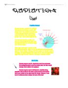
Radiation and Nuclear medicine
Nuclear medicine is a relatively new type of medicine and it incorporates the use of radioactive isotopes of stable elements bound to bodily substances in order to target certain organs and body parts for diagnosis.
Scientists now have the technology to create radioactive forms of , these are called isotopes. Each isotope has a fixed rate of decay which can be characterized by its half-life, a half life is the amount of time it takes for half of the radioactive atoms in a sample to decay to stable elements. Because each isotope decays at a unique and predictable rate, different isotopes are used for a variety of purposes. Some isotopes used and there uses are listed below.
Cesium - 137: Used to treat cancers
Cobalt - 57: Used to help doctors interpret diagnosis scans of organs.
Copper - 67: When injected with monoclonal antibodies into a cancer patient, helps the antibodies bind to and destroy the tumor
Iodine - 131: Used in the diagnosis and treatment of thyroid disorders
Strontium - 85: Used to study bone formation and metabolism.
Xenon - 133: Used for lung ventilation and blood flow studies
Technetium - 99m: Different chemical forms are used for bone, brain, spleen, liver and kidney imaging and also for blood flow studies
Isotopes are ingested and traced in their path through the body, revealing metabolic and biochemical processes extremely accurately. These isotropic "tracers" are currently used for practical diagnosis of disease.
Only certain isotopes are useful however and the way physicians differentiate and chose which isotope to use is to do with their half-lives and their method of decay. If isotopes decay and release alpha particles they cannot be used, as alpha radiation is incredibly ionizing. Beta radiation is not ideal as it cannot penetrate thick skin though gamma radiation which is low ionizing yet high penetrating is only produced with beta radiation. Therefore isotopes which under go beta decay must be used. Isotopes must also have a suitable half-life, half-lives range from 4.5bn years to nano-seconds. Isotopes which are generally used have half-lives of several hours so that it decays quickly, so no bodily damage occurs, but slow enough for a diagnostic picture to be created.
Technetium-99m is the most commonly used radionuclide as it emits gamma radiation and has a half life of 6 hours. Approximately 80% of diagnoses are done with technetium-99m. technetium-99m can be manufactured however by the time it was packaged, reached the hospital and then the patient it would have almost entirely decayed, therefore hospitals are provided with a generator (technetium cow) which contains a parent isotope which decays to form technetium-99m. The parent isotope is molybdenum-99 which can be easily bound to a cartridge for transportation and storage. However technetium-99m cannot be easily bound to the cartridge therefore when it is needed it can be easily washed into another cartridge by sterile water from this lead bound container the isotope can be administered to the patient.
The aim of nuclear medicine is the development of receptor-specific carrier molecule, to which isotopes can be bound, which target specific organs or disease states, and carry technetium-99m or other radionuclides to the sites in the body that want imaging. Once this binding has occurred the substance is termed a radiopharmaceutical and can be administered in three different ways depending on where it must end up. It can be swallowed, injected or inhaled.
The picture is created by a tomography using gamma receptors (photomultipliers) which change the colour of their crystals depending upon the amount of gamma radiation it receives. After this the colour change occurs larger photomultipliers send a signal to a computer where the picture is built up in slices (cross-sectional of a horizontal body) the brighter areas are where there is a higher level of gamma radiation coming from.