There is NO interphase between Meiosis I and Meiosis II
10.1.2: Outline the formation of chiasmata in the process of crossing over.
1. Homologous chromosomes commencing pairing to form a bivalent as they continue to shorten and thicken by coiling.
2. Breakages occur in parallel non-sister chromatids at identical points.
3. Rejoining of non-sister chromatids forms chiasmata.
4. Positions of chiasmata become visible later, as tight pairing of homologous chromosomes ends.
5. When homologous chromosomes move apart in anaphase I, crossing over becomes fully apparent.
10.1.3: Explain how meiosis results in an effectively infinite genetic variety in gametes through crossing over in prophase I and random orientation in metaphase I.
If a homologous pair is denoted as having chromosomes A and B paired together, random orientation during Metaphase I means that in any one cell after meiosis I, the cell could have either chromosome A or B, creating a random orientation of chromosomes in haploid cells that leads to genetic variability.
Added to this is the effect of crossing over during Prophase I, meaning that chromosomes could have any combination of chromosomes A or B, creating an almost infinite genetic variability.
10.1.4: State Mendel’s law of independent assortment
This law states that allele pairs separate independently during the formation of gametes. Therefore, traits are transmitted to offspring independently of one another.
10.1.5: Explain the relationship between Mendel’s law of independent assortment and meiosis.
The relationship between Mendel’s law of independent assortment and meiosis is that the separation of the allele pairs occurs during meiosis. This links to Mendel’s idea that allele pairs separate independently during the formation of gametes.
Theoretical Genetics
4.3.1: Define genotype, phenotype, dominant allele, recessive allele, codominant alleles, locus, homozygous, heterozygous, carrier and test cross.
Genotype - the alleles of an organism
Phenotype - the characteristics of an organism.
Dominant allele - an allele that has the same effect on the phenotype whether it is present in the homozygous or heterozygous state.
Recessive allele - an allele that only has an effect on the phenotype when present in the homozygous state.
Codominant allele - pairs of alleles that both affect the phenotype when present in a heterozygote.
Locus - the particular position on homologous chromosomes of a gene.
Homozygous - Having two identical alleles of a gene
Heterozygous - Having two different alleles of a gene.
Carrier - an individual that has one copy of a recessive allele that causes a genetic disease in individuals that are homozygous for this allele.
Test cross - testing a suspected heterozygote by crossing it with a known homozygous recessive.
4.3.2: Determine the genotypes and phenotypes of the offspring of a monohybrid cross using a Punnet grid.
The grid should be labelled to include parental genotypes, gametes, and both offspring genotype and phenotype.
4.3.3: State that some genes have more than two alleles (multiple alleles).
Some genes have more than two alleles (multiple alleles). Most genes, in fact, have more than two alleles for example ABO blood groups.
4.3.4: Describe ABO blood groups as an example of codominance and multiple alleles.
With multiple alleles, one chooses a single capital letter to represent the locus at which the alleles may occur, and the individual alleles are then represented by a additional single letter (usually capital) in a superscript position. This is done in codominant alleles.
There are 4 blood groups: A, B, O and AB.
This blood group system is determined by combinations of alternative alleles. In each individual, only two of the three alleles exist, but they are inherited as if they were alternative alleles of a pair. However, IA and IB are codominant alleles and both IA and IB are dominant to the recessive Ii.
4.3.5: Explain how the sex chromosomes control gender by referring to the inheritance of X and Y chromosomes in humans.
Females have XX chromosomes.
Males have XY chromosomes.
Just stick these into a punnet grid and show that 50% of boy or girl.
4.3.6: State that some genes are present on the X chromosome and absent from the shorter Y chromosome in humans.
Some genes are present on the X chromosomes and absent from the shorter Y chromosome in humans.
4.3.7: Define sex linkage.
Sex linkage is a special case of linkage occurring when a gene is located on a sex chromosome (usually the X chromosome).
4.3.8: Describe the inheritance of colour blindness and haemophilia as examples of sex linkage.
Both colour blindness and haemophilia are produced by a recessive sex-linked allele on the X chromosome Xb and Xh is the notation for the alleles concerned. The corresponding dominant alleles are XB and XH.
A red-green colour blind person sees green, yellow, orange and red as all the same colour. The conditions afflicts about 8% of males, but only 0.4% of females in the human population. This is because a female with normal colour vision may be homozygous for the normal colour vision allele (XBXB) or she can be heterozygous and a carrier of the colour-blind gene (XBXb) For a female to be colour-blind, but X chromosomes must carry it and therefore be homozygous recessive (XbXb). Males however only have 1 X chromosome and if this X chromosome is affected then they will have colour blindness.
Haemophilia is a disorder in which the blood will not clot normally and they experience continuous, excessive bleeding. There are two types – Type A and Type B.
It is sex linked, the genes controlling the production of the blood proteins concerned are located on the X chromosome. It is caused by a recessive allele. Like colour blindness, it affects the male population considerably more than the female population. For a female to have this condition, they must be homozygous recessive, however this is usually a fatal and the baby does not survive to be born.
Haemophilic gene: Xh.
Normal: XH
4.3.9: State that a human female can be homozygous or heterozygous with respect to sex-linked genes.
A human female can be homozygous or heterozygous with respect to sex-linked genes.
4.3.10: Explain that female carriers are heterozygous for X-linked recessive alleles.
Female carriers are heterozygous for X-linked recessive alleles.
-
A female carrier must be heterozygous as one X chromosome has the dominant allele and the other has the recessive allele XH Xh
- The term carrier can be applied to any individual that is heterozygous for a recessive gene
4.3.11: Predict that the genotypic and phenotypic ratios of offspring of monohybrid crosses involving any of the above patterns of inheritance.
---
4.3.12: Deduce the genotypes and phenotypes of individuals in pedigree charts.
-
Roman numeral identifies generation number
- Arabic numeral identifies individual within a generation, numbering from left to right
- square = male; circle = female
- shaded = affected by genetic disorder or trait
For dominant and recessive alleles, upper-case and lower-case letters, respectively, should be used. Letters representing alleles should be chosen with care to avoid confusion between upper and lower case.
For codominance, the main letter should relate to the gene and the suffix to the allele, both uppercase. For example, red and white codominant flowers colours should be represented as CR and Cw respectively. For sickle-cell anaemia, Hb^ is normal and Hb’ is sickle cell.
Genetic Engineering and Biotechnology
4.4.1: Outline the use of polymerase chain reaction (PCR) to copy and amplify minute quantities of DNA.
Polymerase chain reaction (PCR) is used when a research would like to investigate a certain DNA sequence. This sequence is ‘photocopied’ several times by using enzymes to replicate DNA, thus no living organisms are required.
The DNA is heated to break the hydrogen bonds. Primers are added to start the process of DNA replication. Mixture is cooled and the primers bond to the original, but now single stranded DNA. Nucleotides and a thermostable DNA polymerase are added.
The nucleotides will bond with the ‘exposed’ organic bases of the single stranded DNA then the DNA polymerase will then join them into a DNA strand. Through this, each of the original strands form a new complementary strand.
These strands are heated and separated and will fnction as a template for more DNA strands to be formed. A large amount of identical copies of the original DNA can therefore be made quickly.
4.4.2: State that, in gel electrophoresis, fragments of DNA move in an electric field and are separated according to their size.
In gel electrophoresis, fragments of DNA move in an electric field and are separated according to their size. It involves the separation of DNA, RNA and or other protein molecules through the use of electricity.
Gel electrophoresis is a technique used to separate large molecules, based on their different rates of movement in an electric field cause by a combination of their charge and their size.
4.4.3: State that gel electrophoresis of DNA is used in DNA profiling.
Gel electrophoresis of DNA is used in DNA profiling.
DNA profiling is also known as DNA fingerprinting and is used to compare DNA from different sources without mapping the entire genome. Comparison of DNA from suspect and sample.
4.4.4: Describe the application of DNA profiling to determine paternity and also in forensic investigations.
4.4.5: Analyse DNA profiles to draw conclusions about paternity or forensic investigations.
The outcomes of this analysis could include knowledge of the number of human genes, the location of specific genes, discovery of proteins and their functions and evolutionary relationships.
4.4.6: Outline three outcomes of the sequencing of the complete human genome.
- Improved knowledge about the functions of our genes
- Improved diagnosis of genetic disorders
- Increased potential of gene therapy
- Increased potential to develop drugs that can be used to combat genetic malfunctions.
4.4.7: State that, when genes are transferred between species, the amino acid sequence of polypeptides translated from them is unchanged because the genetic code is universal.
When genes are transferred between species, the amino acid sequence of polypeptides translated from them is unchanged because the genetic code is universal.
Genetic engineering is the deliberate manipulation of genes. Genetic code can be translated from species because the code is universal. This means that for every organism, the same RNA codon codes for the same amino acid in an mRNA strand. As this is possible, it is therefore possible to transfer genetic material from one species to another for example, the human gene for insulin production into a bacterium.
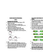
4.4.8: Outline a basic technique for gene transfer involving plasmids, a host cell (bacterium, yeast or other cell), restriction enzymes (endonucleases) and DNA ligase.
Gene transfer involves
- A vector
- Host cell
- Restriction enzymes
- DNA ligase
- The plasmid is removed from the bacterium and the T-DNA is cut by a restriction enzyme
- Foreign DNA is cut by the same enzyme
- The foreign DNA is inserted into the T-DNA of the plasmid
- The plasmid is reinserted into the bacterium
- The bacterium is used to insert theT-DNA carring the foreign gene into the chromosome of a plant cell
- The plant cells are grown in culture
- A plant is generated from a cell clone. All of its cells carry the foreign gene and may express it as a new trait.
The use of E.coli in gene technology is well documented. Most of its DNA is in one circular chromosome, but it also has plasmids (smaller circles of DNA). These plasmids can be removed and cleaved by restriction enzymes at target sequences. DNA fragments from another organism can also be cleaved by the same restriction enzyme, and these pieces can be added to the open plasmid and spliced together by ligase. The recombinant plasmids formed can be inserted into new host cells and cloned.
4.4.9: State two examples of the current uses of genetically modified crops or animals.
Bt corn is genetically modified maize in which a gene from Bt had been incorporated into the maize DNA. This let the plant produce a toxin that makes it resistant to insects. They are grown around the US.
Those who have rice as a major component of their diet may suffer from vitamin A deficiency that can lead to blindness. Rice plants store the vitamin in their leaves, not their grain, however Golden rice is genetically modified by adding genes from daffodils and from a bacterium. This allows the plant to store beta carotene, a precursor of vitamin A in the grains, causing the yellow colour. This is done so that people can meet the recommended intake of vitamin A.
Examples include salt tolerance in tomato plants, synthesis of beta-carotene (vitamin A precursor) in rice, herbicide resistance in crop plants and factor IX (human blood clotting) in sheep milk.
4.4.10: Discuss the potential benefits and possible harmful effects of one example of genetic modification.
BT Corn – contains a gene from the Bacillus thuringiensus which produces a protein that is TOXIC to insects. Found in Europe and US.
Benefits:
- damaged caused by insects are reduced
- Bt corn is slightly more expensive, but the difference is less than on extra application of insecticide
- Non- Bt corn needs to be checked often for signs of insects – therefore less checking of corn.
- less insecticide is used therefore less impact on environment and lower health risks for the workers.
- also seems to reduce infection with fungus so toxins produced by fungus are lowered.
Harmful effects:
- also kill some other insects
- insects may develop resistance to Bt toxin because they are exposed to it all the time
- resistant insects also make Bt spray useless as insecticide. Difficult to prevent pollen from travelling outside the field where the Bt corn is grown. Therefore it can fertilise non-Bt corn and they can’t be sold as ‘organic’ foods or fertilise wild plants and make them less resistant to insects.
4.4.11: Define clone.
Clone - a group of genetically identical organisms or a group of cells derived from a single parent cell.
4.4.12: Outline a technique for cloning using differentiated animal cells.
There are two types of cloning – reproductive and therapeutic cloning.
4.4.13: Discuss the ethical issues of therapeutic cloning in humans.
Therapeutic cloning is the creation of an embryo to supply embryonic stem cells for medical use.
AHL
Dihybrid crosses and gene linkage
10.2.1: Calculate and predict the genotypic and phenotypic ratio of offspring of dihybrid crosses involving unlinked autosomal genes.
Dihybrid cross – YYRR x yyrr.
10.2.2: Distinguish between autosomes and sex chromosomes.
Autosomes are chromosomes which are not sex chromosomes.
Sex chromosomes are those chromosomes which help to determine the sex of an individual.
Humans have 22 pairs of autosomes and 2 sex chromosomes.
Females have XX and males have XY sex chromosomes.
X chromosomes are larger and carry more genes than Y chromosomes.
10.2.3: Explain how crossing over between non-sister chromatids of a homologous pair in prophase I can result in an exchange of alleles.
Crossing over takes place during prophase I. The number of chiasmata may differ. Chiasmata can occur between any non-sister chromatids. It is even possible to have multiple chiasmata on two non-sister chromatids.
The number of different types of gametes produced by random orientation alone is 2n where n = haploid number. Add to this the effect of crossing over and the resulting variation is almost infinite.
(Topic 10.1.3)
10.2.4: Define linkage group.
Linkage group refers to a group of genes whose loci are on the SAME chromosome.
10.2.5: Explain an example of a cross between two linked genes
Example looking at the notation TtBb where it was used for non-linked dihybrid cross.
To represent that it is linked, the two alleles must be represented as vertical pairs
10.2.6: Identify which of the offspring are recombinants in a dihybrid cross involving linked genes.
Recombinants in linked genes are those combinations of genes which the parents DID NOT possess.
Polygenic Inheritance
10.3.1: Define polygenic inheritance
Polygenic inheritance is the inheritance of phenotypes that are determined by the collective effect of several genes.
Discontinuous variation – there is no intermediate form and no overlap between the two phenotypes (i.e. two alleles for tall and dwarf plants.) You either have the characteristic or you don’t. For example, blood groups because you can either have one blood group or another, nothing in between. Controlled by alleles of a SINGLE gene or a SMALL number of genes. Environment has little effect.
Continuous variation – there is a complete range of measurements from on extreme to the other. It is the combined effect of many genes → polygenic inheritance, and is often influenced by environmental factors.
10.3.2: Explain that polygenic inheritance can contribute to continuous variation using two examples, one of which must be human skin colour.
Polygenic inheritance can contribute to continuous variation. Two examples of this is human skin colour and the seed colour of wheat.
Seed colour of grain – this is controlled by 3 genes that interact together to control the phenotype. In this case, for all three of these genes there are only 2 alleles. Despite these limitations, the full range of colours obtained varies from dark red to white, with 5 shades of pink.
Human skin colour – ranges from white, shades of brown to black.
Colour of skin is due to pigment called melanin. Approx 3+ inherited genes control melanin production. The outcome is an almost continuous distribution from very pale (no alleles coding for melanin production) to very dark brown (all alleles for skin colour code for melanin production).