Positron Emission Tomography (PET)
- Positron Emission Tomography (PET) scans detect special radioactively labelled tracers which are injected into a patient's body before the imaging procedure starts.
- PET scans can be used to accurately monitor brain activity while a patient's memory and cognition are being tested.
- PET scans can determine brain activity and function by measuring differences in blood flow and the usage of glucose (sugar), both of which increase when an area of the brain is active.
- PET scans provide information about brain function and activity as opposed to brain structure, and are more typically used in research.
- The scans are made by injecting the patient with a form of sugar that has been altered to carry a weak, short-lived radioactive element. The sugar hits the bloodstream and flows to the brain, which needs huge amounts of energy to keep all its nerve cells running.
- The most active areas of the brain need the most sugar -- while damaged and less active areas need much less. By detecting the weak radiation signal from the sugar molecules as they travel throughout the brain, PET scanners can make a picture of brain cell activity. The resulting scans show the level of activity using a scale of colours; red and orange for high activity, and blue and purple for low.
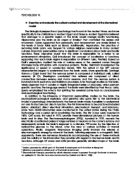
- Researchers from the New York University School of Medicine have developed a brain-scan-based computer programme that quickly and accurately measures metabolic activity in the hippocampus. Using PET scans and the computer programme the researchers showed that in the early stages of Alzheimer’s disease there is a reduction in brain metabolism in the hippocampus.
- In a longitudinal study they followed a sample of 53 normal and healthy participants – some for 9 years and others for as long as 24 years. They found that individuals who showed early signs of reduced metabolism in the hippocampus were associated with later development of Alzheimer’s disease.