Secondary Structure
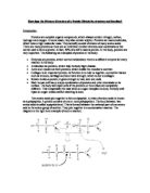
There are two types of secondary structure in proteins, the helix and the pleated sheet. The attraction of the R groups within the same chain can cause the chain to twist into a "right handed" coil. This helix is held together by hydrogen bonds between the hydrogen and oxygen atoms of the amino acid backbone (amino groups and carboxyl groups). Below is a diagram to show how these bonds occur.
The β pleated sheet is formed when hydrogen bonds occur between the C=O and NH groups of one chain and the C=O and NH of the next chain. Below is a picture to show how to parallel chains of amino acids can form hydrogen bonds between each other.
Important things to remember concerning the secondary structure are:
- The secondary structure is determined by the primary structure. Certain amino acids easily form helices and partial sequences of peptides that contain a large amount of these amino acids will most likely form such helices. Sequences that don't will not form them. The same could be said for the beta-sheet.
- Secondary structures do not usually describe the structure of the entire protein but of smaller, local regions of the peptide chain.
- Helices and sheets are held together by numerous hydrogen bonds between atoms that flank the peptide bond (remember, R-groups are not directly involved in the formation of secondary structure). While hydrogen bonds are weak interactions, the large number of hydrogen bonds that form in helices (or sheets) make the structure very stable.
- Since the helices and sheets are held together by hydrogen bonds, any condition that may interfere with the formation of these bonds can disrupt and destroy the structure. Examples of factors that interfere with secondary structure are heat (heat increases the vibrations of atoms thus breaking hydrogen bonds), changes in pH, and high ionic strength (high concentration of H+ and of ions in general will interfere with the formation of hydrogen bonds). This effect on the protein secondary structure is part of the reason why the function of proteins is affected by temperature, ionic strength and pH.
Here is a picture of the two different structures, some proteins can have an alpha helix and a beta pleated sheet in the same molecule.
The secondary structure is also important as it can also have a bearing upon the function of the protein. For example, collagen is a triple helix structure. This means that it is made of three helical shapes wrapped around each other. This means it is very strong and is ideal for connective tissues such as tendons in the body.
Tertiary Structure
The tertiary structure of a protein is a description of the complex and irregular folding of the peptide chain in three dimensions. It is essentially a picture of what the shape of the entire protein actually looks like. There are two general tertiary structures that a protein can have, globular and fibrous.
Globular proteins are characterized as generally having:
- A variety of different kinds of secondary structure
- A spherical shape
- A good water solubility
- A catalytic/regulatory/transport role, i.e., a dynamic metabolic function.
Fibrous Proteins are characterized as generally having:
- One dominating kind of secondary structure (i.e., collagen helix in collagen
- A long narrow (rod-like) structure.
- A low water solubility
- A role in determining tissue/cellular structure
There are five variables that can affect the tertiary structure. They are:
- Hydrophobic interactions: These cause the structure to fold as the protein shields the hydrophobic side-groups from the aqueous surroundings
- Van der waals forces: There are weak inter-molecular forces between the chains.
- Hydrogen bonds: These occur between the two R groups, and can cause the chain to bend.
- Disulphide bonds: covalent S-S bonds between two cysteine amino acids, which are strong.
- Ionic bonds: Bonds between positive amine and negative carboxyl.
Quaternary Structure
The quaternary structure of a protein describes the interactions between different peptide chains that make up the protein. Some proteins (such as haemoglobin) have more than one peptide chain (these are multimeric proteins). The manner in which these chains fit together is the quaternary structure. Obviously, if a protein is made up of only one chain (monomeric), there is no quaternary structure for that protein. The forces that hold different chains together are the same that hold the tertiary structure together, hydrogen bonding between polar R-groups, ionic bonds between charged R-groups, hydrophobic interactions between non-polar R-groups, and disulfide bonds. The figure below shows the structure of haemoglobin, a protein that has four subunits. Each subunit is identified with a different colour.
Collagen
Collagen is a major structural protein, forming molecular cables that strengthen the tendons and vast, resilient sheets that support the skin and internal organs. Bones and teeth are made by adding mineral crystals to collagen. Collagen provides structure to our bodies, protecting and supporting the softer tissues and connecting them with the skeleton. Collagen is composed of three chains, wound together in a tight triple helix. The diagram shown is a small section of the collagen chain; in reality each chain is over 1400 amino acids long and only about 20 are shown here. A repeated sequence of three amino acids forms this sturdy structure. Every third amino acid is glycine, a small amino acid that fits perfectly inside the helix. Many of the remaining positions in the chain are filled by two unexpected amino acids: proline and a modified version of proline, hydroxyproline.
The body makes many different kinds of collagen, which form long ropes and tough sheets that are used for structural support in mature animals and as pathways for cellular movement during development. All contain a long stretch of triple helix connected to different types of ends. The simplest is a long triple helix, with blunt ends. These type I collagen molecules associate side-by-side, like fibres in a rope, to form tough fibrils. These fibrils crisscross the space between nearly every one of our cells. This prevents the collagen from being stretched; the fibrils act as covalent bonds, which hold the structure together.
Collagen is made up of long-stranded molecules, called tropocollagen, organised in small bundles (microfibrils and fibrils) so that each strand has a large overlap with others. The tropocollagen molecules cross-link covalently to each other using lysine side chains, these cross-links are unusual and occur only in collagen and elastin, a related protein.
The fibrils may contain millions of tropocollagen strands. They run parallel along the length of a tendon. The secondary structure of tropocollagen is unusual. Each strand of tropocollagen is made from three individual polypeptide chains. When a muscle contracts, collagen fibres in the connecting tendons have the job of passing the effect on to the skeleton to bring about some movement. It is important that the collagen fibres in the tendons are strong and do not stretch significantly. A collagen fibre of 1 mm diameter can support a mass of at least 10 kg before it breaks. When collagen is pulled in a direction parallel to its fibres it is the covalent bonds that provide the strength and resistance.
Insulin
Insulin is constructed of two peptide chains, referred to as A and B, which are held together by two disulfide bonds between the cysteine amino acids. Due to there being many amino acids in insulin that contain carbon in the centre of the protein, insulin has a hydrophobic centre, which is then surrounded on the outside by hydrophilic, charged amino acids. This arrangement provides insulin with a very stable protein structure
Although the amino acid sequence of insulin varies among species, certain segments of the molecule are the same, including the positions of the three disulfide bonds, both ends of the A chain and the C-terminal residues of the B chain. These similarities in the amino acid sequence of insulin lead to a three dimensional conformation of insulin that is very similar among species, and insulin from one animal is very likely to work in other species. For example, pig insulin has been widely used to treat human patients.
Myoglobin
Myoglobin is a small, bright red protein. It is very common in muscle cells, and gives meat much of its red colour. Its job is to store oxygen, for use when muscles are hard at work. The structure contains one protein chain, a haem group (with a water molecule bound to the iron), and a sulphate ion. There are several things to look for in this structure below. The protein chain is composed of spring-shaped alpha helices, linked together by short loops. The chain surrounds the flat haem group. At the centre, you can see the iron atom, surrounded by four blue nitrogen atoms.
The first picture shows only a set of thin tubes to represent the protein chain, and the oxygen is easily seen. But when all of the atoms in the protein are shown in the second picture, the oxygen disappears, buried inside the protein. To allow the oxygen to move in and out of the molecule is done very easily. In reality, myoglobin (and all other proteins) are constantly in motion, performing small flexing and breathing motions. Temporary openings constantly appear and disappear, allowing oxygen in and out.
Haemoglobin
Haemoglobin is found in red blood cells. The haemoglobin molecule is a tetramer consisting of 4 polypeptide chains, known as globins, which are usually:
- 2 alpha chains that are each 141 amino acids long
- 2 beta chains that are each 146 amino acids long
Attached to each chain is an iron-containing molecule known as a haem group.
Oxygen binding at the four haem sites in haemoglobin does not happen simultaneously. Once the first heme binds oxygen, it introduces small changes in the structure of the corresponding protein chain. These changes nudge the neighbouring chains into a different shape, making them bind oxygen more easily. Thus, it is difficult to add the first oxygen molecule, but binding the second, a third and fourth oxygen molecule gets progressively easier and easier. This provides a great advantage in haemoglobin function. When blood is in the lungs, where there is a lot of oxygen, it easily binds to the first subunit and then quickly fills up the remaining ones. Then, as blood circulates through the body, the oxygen level drops while that of carbon dioxide increases. In this environment, haemoglobin releases its oxygen. As soon as the first oxygen molecule drops off, the protein starts changing its shape. This prompts the remaining three oxygens to be quickly released. In this way, haemoglobin picks up the largest possible load of oxygen in the lungs, and delivers all of it where and when needed.
The genes for the protein chains of haemoglobin show small differences with different people, so the amino acid sequence of haemoglobin is slightly different from person to person. In most cases the changes do not affect protein function and are often not even noticed. However, in some cases these different amino acids lead to major structural changes. One such example is that of the sickle cell haemoglobin, where glutamate 6 in the beta chain is mutated to valine. This change allows the deoxygenated form of the haemoglobin to stick to each other producing stiff fibres of haemoglobin inside red blood cells. This in turn deforms the red blood cell, which is normally a smooth disk shape, into a C or sickle shape. The distorted cells are fragile and often rupture, leading to loss of haemoglobin.
Bibliography
Mrothery website: http://www.mrothery.co.uk/
Protein structure websites:
http://www.blc.arizona.edu/courses/181gh/rick/biomolecules/protein.html
http://old.jccc.net/~pdecell/biochemistry/protstruc.html
http://webhost.bridgew.edu/fgorga/proteins/proteins.htm
http://www.piercenet.com/Proteomics/browse.cfm?fldID=FE2F8423-A949-11D5-9E2A-00508BD9167A
Protein Data bank: http://www.rcsb.org/pdb/index.html
Biological science 1 textbook