Firstly, the female patient is subjected to COS, to obtain many oocytes. Biopsies are then carried out at several different stages during the process for diagnosing whether the embryos are at risk from the disease; on fertilised and unfertilised oocytes, at day 3- cleavage stage embryos, and on blastocysts. When performing tests on the fertilised and unfertilised oocytes, polar bodies are extracted from the mother. The first biopsy is performed on the unfertilised 1st polar body, whereas the second biopsy is performed on the 2nd polar body after IVF and the successful fertilisation of the oocyte. The biopsy at day 3 is performed on the embryo when it is at the 8 cell stage, and the final biopsy is carried out on blastocysts. The use of blastocysts increases the sample size that is used in the techniques to determine whether the embryo has the mutated genes or not; this increases the accuracy of the whole process.
The techniques used in the determination of whether an embryo is a carrier of the disease are PCR and Fluorescent In Situ Hybridisation (FISH).
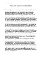
An example of the graphical representation of FISH (http://www.frontiersin.org/neurogenomics/10.3389/fnins.2010.00165/full)
The use of PCR is mainly to diagnose monogenic conditions, such as cystic fibrosis, sickle cell disease, and Huntingtons disease (Strode. A, 2011). The disorders are all autosomal recessive, autosomal dominant and X-linked diseases and are all due to the mutation of a single gene. The use of PCR to diagnose these diseases had led to some dispute over the accuracy of the results, due to the fact that the DNA must be amplified from a single strand of DNA, which is the smallest possible template that can be used. Because of this, modification of the PCR system must be undertaken to ensure the correct amplification (Rychlik. W, 1990). As this process is easily contaminated, and subject to misdiagnosis, a study was carried out to try to quantify the errors that are made. (Lewis. CM, 2000)
The FISH technique uses DNA probes that have a fluorochrome attached and is used to diagnose chromosomal abnormalities, like aneuploidy screening; which is when the cell has an abnormal number of chromosomes. This process has also been used to sex the embryo. This only happens when there is no procedure for PCR for a given X-linked disease. Doubt has been cast over the accuracy of this procedure due to testing of some embryos after they were classed as unfit by the FISH test. Some of these embryos were found to be normal, healthy embryos leading to a decreased chance of pregnancy due to a lower number of viable embryos (Staessen. C, 2004).
Once these processes have been performed, implantation can take place. This usually occurs on day 3 or 5 after fertilisation takes place. In Europe, only one embryo may be transferred, reducing the risk of multiple pregnancies, although this does also lower the probability of a successful pregnancy occurring. Other viable embryos may undergo cryopreservation, to ensure that they remain viable for a long period of time.
As the aim of PGD is to identify mutated genes in an embryo, and then discard of that embryo, the chance of the child then developing that disease is effectively eradiated (although the effectiveness is not 100%). This, therefore, means that the inherited disease has been removed from the family line.
This process has also been used in Human Leukocyte Typing on embryos. The reason for this is so that when the child is born, it may provide cordial blood stem cells that can be used to treat a sibling that already has a disease. These children are sometimes called ‘Saviour Siblings’. These children have caused the biggest controversy, as it is argued that the child is exploited and only born for the purpose of saving another (Strode. A, 2011).
The first baby born in the UK using PGD was screened for a defective BRCA1 gene, which is a gene associated with breast and ovarian cancer. The father of the child had a long family history of breast cancer, with all his immediate female family developing the disease. The gene that caused this was identified as the defective BRCA1 gene. PGD was then used to screen the embryos for this defect, and only the healthy ones were selected and then implanted. The pregnancy resulted in a healthy baby girl that was free from the defective gene and free from the disease (http://www.dailymail.co.uk/health/article-1110244/Britains-cancer-free-designer-baby-born-screened-deadly-gene.html).
Also, on rare occasions, PGD has been used for the opposite effect. A deaf lesbian couple wished to have a child that shared their disability, so embryo screening was used to ensure this happened (http://www.newscientist.com/blog/shortsharpscience/2006/09/designer-deafness.html). This caused an ethical storm.
PGD is still a controversial topic, with human rights groups campaigning that this could lead to a larger social divide. As PGD, coupled with IVF, is an expensive procedure, it is argued that only the rich will be able to afford to have this procedure. It is also argued that a race of ‘genetically enhanced humans’ with increased desirable traits (intelligence, height etc.) would look down on those that could not afford PGD, and still suffered from inherited diseases.
However, PGD is more ethical than prior techniques used for screening like prenatal screening. This process involved taking a biopsy after the embryo has been implanted and if the embryo was found to contain the defective disease, it would be aborted. Whereas with PGD, no abortion needs to occur.
In conclusion, the creation of designer babies is not how the public and the media perceive it to be. PGD is used to select embryos based on a purely medical basis, like the eradication of genetic disease, not to tailor the baby’s specific appearance to the parent’s wishes. In the UK, where the creation of designer babies is tightly regulated by the Human Fertility and Embryology Authority and each case is discussed separately, the ‘genetically enhanced humans’ remain in the far future. The technique used in the creation of designer babies remains solely embryo selection, and not gene therapy.
Word Count – 1345
Bibliography
Handyside. AH, K. E. (1990). Pregnancies from biopsied human preimplantation embryos sexed by Y-specific DNA amplification. Nature , 768-70.
http://www.dailymail.co.uk/health/article-1110244/Britains-cancer-free-designer-baby-born-screened-deadly-gene.html. (n.d.).
http://www.frontiersin.org/neurogenomics/10.3389/fnins.2010.00165/full. (n.d.).
http://www.newscientist.com/blog/shortsharpscience/2006/09/designer-deafness.html. (n.d.).
Lewis. CM, P. T. (2000). Controlling misdiagnosis errors in preimplantation genetic diagnosis: a comprehensive model encompassing extrinsic and intrinsic sources of error. Hum. Reprod , 43-50.
Rychlik. W, S. W. (1990). Optimization of the annealing temperature for DNA amplification in vitro. Nucleic Acids Res , 6409-12.
Staessen. C, C. M. (2004). Comparison of blastocyst transfer with or without preimplantation genetic diagnosis for aneuploidy screening in couples with advanced maternal age: a prospective randomized controlled trial. Hum. Reprod , 2849-58.
Strode. A, S. S. (2011). Preimplantation diagnosis to create 'saviour siblings': A critical discussion of the current and future legal frameworks in South Africa. S Afr Med J. , 21.