CLBP is continually seen in the clinical setting and although research into this area shows mixed results, some evidence suggests proprioceptive deficits do exist. Research into this area will help our understanding of CLBP and determine how it is treated. This study has used patients with diagnosed CLBP on a piece of equipment that is used in clinical practice. It will therefore give results that are easily transferable to the clinical setting. It aims to determine whether proprioceptive deficits are present in the spine by looking at repositioning error and proprioceptive awareness with and without visual feedback.
Method
Subject Selection
Ten participants, six females and four males, with a mean age of 54.8(s.d 7.9.) were consented to participate in the study. All the participants have had low back pain for over three months and were attending a rehabilitation programme for CLBP. All were referred via a chartered physiotherapist and completed an Oswestry Disability Index (ODI) questionnaire. Details of the patients are shown in Table one, although some data was unobtainable. Exclusion criteria
included any history of lower limb injuries or complaints and any conditions that could affect their balance. Ethical
approval was authorised by the Oxfordshire LRECcommittee and patients were informed that they were free to withdraw from the study at any time.
Apparatus
The Balance Performance Monitor (BPM) has a feedback unit that is attached to two footplates and connected to a PC containing the SMS software for analysing the results. It records the sway area, sway path and mean percentage weight distribution in standing over thirty seconds. The feedback unit was turned to silent and placed away from the participants view so that they would not have any visual feedback.
Table One
Patient Demographics
Procedure
Experiment 1
Each participant’s height was taken and entered into the SMS software so as to determine the distance between the footplates to gain each individual’s correct stance. The participants removed their footwear and stood on the footplates with arms crossed to opposite shoulders. The examiner placed the feet as accurately as possible on the footplates. The participant focused on an eye level marker for thirty seconds while the BPM recorded the sway area and sway path. This was repeated for a further thirty seconds with eyes closed while the same data was collected.
Experiment 2
A metre ruler was placed on the wall to act as a measuring marker and a digital
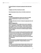
camera was placed 125cms away from the footplates at right angles so as to measure joint position. A target board was placed within a comfortable reaching position, for each individual participant, in front of them. The participants’ top and footwear were removed and they stood on the footplates as in experiment one. C7, T12 and L4 were palpated and a marker applied to the spinous processes (see fig one).
The BPM recorded their mean percentage weight distribution, sway path and sway area for thirty seconds. When instructed the participant leant forward to touch the three marks on the target board with their right hand from right to left and returned to their standing position. The same data was recorded for another thirty seconds. This was repeated but a photograph was taken before they leant forward and another when they returned to their standing position so to determine whether there was any repositioning error. This was calculated by taking a standard marker from the metre ruler and measuring in mms the distance to each marker on the spine and calculating the difference between the two photographs.
Standing was chosen as it is a functional position that is most commonly adopted throughout our daily living. However these experiments are not a true measure of spinal proprioception as feedback will also be given through other joints including the hip, knees and soles of the feet.
Data Analysis
The Wilcoxon paired significance test was used to determine significance of sway area and sway path with eyes open and closed. The same test was used for repositioning error and was calculated via the SPSS computer software, where the
alpha level was set at p<0.05. This test was used as the data is non parametric and related. Spearmans Rho was used as a correlation test between ODI and sway
area with eyes closed with the p value set at the same alpha level.
Fig 1
Example patient for experiment two
Results
The results for the eyes open / eyes closed test are summarised in Table two. As hypothesised the sway area significantly increased with eyes closed (p<0.007) as did the sway path (p<0.005) using the Wilcoxon paired significance test with a p value of p<0.05. Table three summarises
the means and standard deviations of percentage mean balance (weight distribution), sway area and sway path before and after repositioning. As shown, there was not a statistical significance between these measurements before and after repositioning. The mean balance does not give a true reflection as seen in Fig two where it is clear that on repositioning, each patient alters their stance and centre of pressure. Both sway area and sway path increase on repositioning and it appears that these patients have an increased sway area compared to sway path. Repositioning error of C7, T10 and L4 is shown in Table Four but all measurements were insignificant, C7 being p<0.796, T10 p<0.230 and L4 p<0.167 using the Wilcoxon paired significance test with a p value of p<0.05. Fig three examines the relationship between sway area with eyes closed compared to the patients Oswestry Disability Index. Spearmans Rho correlation test was used with the same p value. The correlation coefficient was 0.419 and was therefore insignificant with a p value of p<0.301.
Table Two
Sway area and sway path results for eyes open/closed
Table Three
Mean balance (MB), sway area (SA) and sway path (SP) results before (A) and after (B) repositioning
(%R = % weight distribution to the right)
Table Four
Re-positioning Error
Fig Two
Mean Balance (%) before (a) and after (b) repositioning
Fig Three
Scattergraph comparing ODI and sway area with eyes closed.
Discussion
Experiment one has shown that by removing vision sway area and path significantly increase showing a decrease in postural and righting reactions. Table two shows that sway area is affected more than sway path suggesting that small postural adjustments are not being made due to decreased proprioceptive awareness and it is only when larger movements are made that these patients are aware of their joint position and correct it. Ekdahl (1989) discovered that normal subjects have a higher sway path compared to sway area suggesting they are able to make postural adjustments faster. These postural reactions are necessary to maintain equilibrium and balance, and require synergistic contraction of flexors and extensors with precise timing. It can therefore be seen that balance and posture are intimately related (Trew, 2001). By removing vision, balance is compromised leading to increased postural awareness and is therefore a valid variable for measuring proprioception.
The patients had varied levels of impairment due to back pain as shown on the Oswestry disability index (Table one) and were taken from different stages of the back pain classes which may explain the unusually high standard deviation of sway area with eyes closed. Fig three shows a general correlation between patients’ ODI and their sway area with eyes closed. These variables were chosen as sway area
is most relevant to CLBP patients and the ODI shows how the patients varied in their levels of impairment. A general correlation can be seen, where an increase in ODI leads to an increase in sway area and therefore the more compromising the back dysfunction, the more likely they are to have proprioceptive deficits. As can be seen in fig three there are exceptions to the rule but perhaps a larger sample would show a stronger trend. The ODI could perhaps be a good indicator of proprioceptive deficits and needs to be researched further.
The standing position was chosen as it is functional, however afferent information from the lower limbs may have provided proprioceptive feedback and therefore it is not a true measure of propriception in the lumbar spine. However the only variable changed in this experiment was the removal of vision thereby increasing its validity, and is consistent with Nies (1991) and Mientjes (1999) findings where sway area and path increased under the same condition. Despite Mientjes (1999) study only involving eight patients, limiting its generalisation, there is no research to negate these findings. It is clear that visual feedback plays a vital role in joint awareness. Sackley (1993) found visual feedback training to be effective in stroke patients, who improved their weight distribution over their rehabilitation program, however there is little research into CLBP and visual feedback. Further research needs to be conducted based on this study’s results but it is also important to note that vision is only one aspect of proprioception, and sensory feedback and internal vestibular systems also have proprioceptive roles. Nies (1991) looked at sensory feedback by using an unstable base of support and compared keeping the head still to turning the head for the vestibular system. When these systems were challenged, body sway again increased. This study compared healthy subjects to low back pain patients and found significant differences for sway area, however all the healthy subjects were physiotherapists and physiotherapy students which may have biased the results.
The second experiment focused on repositioning error. Fig two clearly shows that each patient had a preference to weight bear on one foot more than the other, which is consistent with normal subjects (Sackley 1991) who tend to favour one leg by up to 12%. This study showed a maximum of 11.5% asymmetry to the right as shown in fig two suggesting that weight bearing patterns remain consistent between healthy and CLBP subjects, although a larger sample size may show different results. Table three shows that on repositioning, sway area and path increase, although not significantly. A possible explanation for this could be that on repositioning the patients need time to adjust to a comfortable position or that fatigue sets in on the second measurement. The standard deviation remains high for sway area and path showing the different levels of back pain due to the results being inconsistent, and the need to have a larger sample size. Ekdahl (1989) discovered that postural sway increases with age in healthy subjects and therefore the wide range of results may be due to this as the ages ranged from forty two to sixty six.
Although the repositioning error was not significant, L4 showed increased movement. As all the subjects had low back pain, a possible explanation for these results is that receptors for proprioception may be dysfunctional at the lumbar spine and there is some compensation in the thoracic spine as a similar significance level was found here. This suggests proprioceptive deficits may occur globally opposed to segmentally (O’Sullivan 2003). It is unclear whether this is due to the back injury itself or the inactivity caused by the back injury. C7 appears to have little relevance in this study. It is important to note that true repositioning error is difficult to measure in the lumbar spine. It has complex three dimensional movements and this method was unable to record any rotation of the spine. Along with possible proprioceptive deficits all these patients had experienced pain due to nocioceptive input in the lumbar spine. To avoid mechanical irritation leading to nocioceptive input the target board was positioned a comfortable distance away so as to gain a better insight into proprioceptive dysfunction.
Research results vary in this area, often depending on the method employed. O’Sullivan (2003) used the 3Space Fastrack and found significant differences in repositioning error between patients with lumbosacral instability and healthy subjects. This is a complex piece of equipment which requires extensive training but is accurate and more sensitive than the method employed in this study. Newcomer (2000) and Gill (1998) looked at reproducing predetermined target positions and identified proprioceptive deficits, although significance was not found. Again this may be due to a less sensitive method as proprioceptive deficits in peripheral joints may affect spinal position. Gill (1998) also used a chest and pelvis harness which offers a large amount of sensory input, decreasing its validity. Force plates have been commonly used (Nies 1991, 1998, Mientjes 1998) also confirming an increase in postural sway in low back pain patients, however the BPM is used in practice and therefore more clinically relevant.
The BPM cannot measure joint position but has been deemed a valid piece of equipment by Haas et al (2000) when looking at symmetry, but not necessarily when looking at the steadiness aspect of postural control. It has been recognised as being reliable (Haas et al, 1998, Hinman et al, 1997) but they recommend that the mean of three measurements be taken to increase the reliability. Due to time constraints this was not feasible in this study. It should also be noted that Hinman (1997) and Haas (1998) studies’ were carried out on healthy participants and therefore caution should be taken when generalising to the patient population. The BPM has been used as an adjunct to physiotherapy for retraining of balance for stroke patients and has been found effective in providing feedback by Sackley (1992,1993), which offers support for its clinical use.
Although this is a small study it provides interesting background to CLBP and proprioception. It suggests that proprioceptive deficits do exist but perhaps more so in some patients. This may be due to certain low back dysfunctions but as diagnosis of certain conditions is difficult to reach, assessments of each patient should be taken individually and a program devised specifically, incorporating proprioceptive exercises as appropriate. This study has its limitations in terms of its sample size and quality of research and therefore provides a starting point for further research. Research looking at integrating visual feedback for training purposes would be beneficial to determine its clinical use along with continued research into finding the best method for measuring proprioception.
Conclusion
Proprioceptive deficits have been identified in patients with CLBP. This has been significantly shown by a decrease in joint position sense when vision is removed in standing. Although statistically insignificant, repositioning error is seen in the lumbar spine and prompts further research into this area to find a suitable method to measure proprioception and whether specific proprioceptive training is beneficial to patients with CLBP.
REFERENCES
Allison G, Fukushima S. (2003) Estimating three dimensional spinal repositioning error: the impact of range, posture, and number of trials. Spine 28 (22) 2510-2516
Ekdahl C, Andersson S. (1989) Standing balance in rheumatoid arthritis. Scandinavian Journal of Rheumatology 18 33-42
Acknowledgments
I would like to thank Karen Barker for her help during data collection and continued support, along with the patients used in this study.
Ekdahl C, Jarnlo G, Andersson S. (1989) Standing balance in healthy subjects. Scandinavian Journal of Rehabilitative Medicine 21 187-195
Gill KP, Callaghan MJ. (1998) The measurement of lumbar proprioception inindividuals with and without low back pain. Spine 23 (3) 371-377
Haas B and Burden A. (2000) Validity of weight distribution and sway measurements of the balance performance monitor. Physiotherapy Research International 5 (1) 19-32
Haas B and Whitmarsh T. (1998) Inter and intra tester reliability of the balance performance monitor in a non patient population. Physiotherapy Research International 3 (2) 135-145
Hinman M R. (1997) Validity and reliability of measures obtained from the balance performance monitor during quiet standing. Physiotherapy Nov 1, 83 (11) 579-581.
Liu-Ambrose T, Taunton JE, MacIntyre D, McConkey P, Khan KM. (2003) The effects of proprioceptive training on the neuromuscular function of the ACL reconstructed knee: a randomised clinical trial. Scandinavian Journal of Medicine and Science in Sports 13 (2) 115-123
Mientjes MI, Frank JS. (1999) Balance in chronic low back pain patients compared to healthy people under various conditions in upright standing. Clinical Biomechanics 14 (10) 710-716
Newcomer K L, Laskowski ER, Yu B, Larson DR, An K. (2000) Comparing trunk repositioning error in subjects with chronic low back pain and control subjects. Spine 25 (2) 245-250.
Nies N, Sinnot P. (1991) Variations in balance and body sway in middle aged adults. Subjects with healthy backs compared with subjects with low back dysfunction. Spine 16 (3) 325-330
O’Sullivan PB, Burnett A, Floyd AN et al. (2003) Lumbar repositioning in a specific low back pain population. Spine 28 (10) 1074-1079
Sackley C M, Baguley B I, Gent S, Hodgson P. (1992) The use of a balance performance monitor in the treatment of weight bearing and weight transference problems after stroke. Physiotherapy 78 (12) 907-921
Sackley C M, Baguley B I. (1993) Visual feedback after stroke with the balance performance monitor: two single case studies. Clinical Rehabilitation 7 189-195.
Sackley C M, Lincoln N B. (1991) Weight distribution and postural sway in healthy adults. Clinical rehabilitation 5 181-186
Trew M, Everett T. (2001) Human Movement: an introductory text. 4th Ed. London:Churchill Livingstone. p 229
Willems T, Witvrouw E, Verstuyft J, Vaes P, De Clercq D. (2002) Proprioception and muscle strength in subjects with a history of ankle strains and chronic instability. Journal of athletic training 37 (4) 487-493