The cardiac cycle is divided into two major phases, systole, which is when the ventricles contract and blood is released from the heart, and diastole, which is when the ventricles relax and blood flows into the heart. During exercise, skeletal muscle tissue requires more nutrients than at resting state, so the blood vessels dilate (vasodilation), to allow more blood to flow to and from the tissues, maximising the nutrients available for the tissue. The heart rate also increases to account for the increased arterial volume. In athletes, the cardiac output can increase from 5L/minute to 35L/minute with 77% of this flowing to skeletal muscle tissue (as opposed to the 20% at rest) [3]. This aim of this experiment was to test the effect of a short period of mild exercise on the heart rate, systolic blood pressure and diastolic blood pressure in a group of 6 young healthy subjects.
Methods
Six university students who had given their legal consent were involved in this experiment to test heart rate, systolic blood pressure and diastolic blood pressure at various points during an exercise session. Blood pressure (systolic and diastolic) and heart rate was taken and recorded using an accurate heart rate/blood pressure monitor. These values were called the ‘Resting’ rates and corresponded to heart rate and blood pressure at normal resting conditions. This was followed by a mild 5 minute session on an exercise bike at a steady pace of around 15-17km/h. It was made sure that each subject was cycling at a comfortable pace and resistance as this experiment was to test heart rate and blood pressure during a short period of mild exercise. Immediately after this session, heart rate and systolic and diastolic blood pressure were measured and recorded again, using the same monitoring equipment. This task was carried out as quickly as possible after the cycling session as heart rate and blood pressure starts to fall immediately. A 10 minute resting period followed the cycling session as this was agreed to be sufficient time for the heart rate and blood pressure to stabilise. During the resting period, the subject was required to sit down in a relaxed manner and avoid as much exertion as possible.
The data gathered during this experiment was statistically analysed using a statistical software program. The means of each set of results were calculated, as were the standard errors of the means. A series of paired T-tests were then carried out using various sets of results. The significance of these T-tests are explained in the results section.
Results
Heart Rate
The mean resting heart rate in the human subjects was 89.17±3.89 beats per minute (n=6). After a period of 5 minutes on an exercise bike, the mean heart rate increased to 127±7.78 beats per minute (n=6), and increase of ~42% (P<0.005) (n=6) (Fig.1). After a 10 minute period of relaxation (avoiding all possible physical exertion), the mean heart rate decreased to 92.17±8.20 beats per minute (P<0.015) (n=6) (Fig.1).
Figure 1: Effect of exercise on heart rate. Heart rate at resting state, after 5 minutes on a cycle machine at 15-17km/h and after 10 minutes of recovery time. Data represent mean ± standard error measurement; n=6; *P<0.005 rest heart rate vs. exercise heart rate; †P<0.015 exercise heart rate vs. after exercise heart rate.
Systolic and Diastolic Blood Pressure
The mean systolic blood pressure in the resting state was 124.33±8.29 mmHg (n=6). The mean systolic blood pressure increased after the 5 minute exercise period to 142±13.1 mmHg (P<0.021) (n=6). The mean systolic blood pressure decreased after the 10 minute rest period to 116.33±5.45 mmHg. The mean diastolic blood pressure at the resting state was 75.83±5.72 mmHg. After the 5 minute exercise period, there was not a significant difference in mean diastolic blood pressure, 70.50±8.78 mmHg. After the 10 minute recovery period, there was also not a significant difference in mean diastolic blood pressure, 75.67±3.64 mmHg.
Figure 2: Effect of exercise on blood pressure (systolic and diastolic). Systolic and diastolic blood pressure at resting state, after 5 minutes on a cycle machine at 15-17km/h and after 10 minutes of recovery time with no physical exertion. Data represent mean ± standard error measurement; n=6; *(P<0.021) rest systolic blood pressure (mmHg) vs. exercise systolic pressure (mmHg).
Discussion
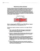
The findings of this experiment conclude that on average, the blood pressures in the sample group in the optimum or normal range. The optimum blood pressure is <120 mmHg for systolic blood pressure and <80 mmHg for diastolic blood pressure. The normal range for systolic blood pressure was 120-139 mmHg and the normal range for diastolic blood pressure was 80-89 mmHg. So the sample group had, on average, and normal systolic blood pressure and a diastolic blood pressure within the optimum level [4].
The heart rate increases during exercise, essentially to meet the bodies demand (in particular skeletal muscle) for nutrients like oxygen. Vasodilation occurs during exercise which is when the blood vessels relax and become wider and thus able to hold and circulate more blood. In order to fill these blood vessels the heart has to pump more blood around the body and process more blood via the lungs to oxygenate it (which is why breathing becomes increased). The only way the heart can process more blood is if it beats faster – to meet the body’s demands [3]. Because there can only be a small increase in stroke volume (due to the heart capacity), there has to be a sharp increase in heart rate to increase the blood flow. The increase in heart rate is caused by decreased parasympathetic nervous activity to the SA node and increased sympathetic activity. These effects are caused by the activity of arterial baroreceptors and mechanoreceptors which act upon the medullary cardiovascular center which in turn decreases parasympathetic activity and increases sympathetic nervous activity [3]. The results obtained during the experiment were conclusive with other sources.
During the experiment, systolic blood pressure increased, and there was a slight decrease in diastolic blood pressure. This is consistent with other research literature [4]. This slight decrease can be explained by the vasodilation of the arteries during exercise, so the expansion of the artery may lower blood pressure during the diastolic phase [4]. The mean arterial blood pressure can be calculated by multiplying the cardiac output (the volume of blood which is pumped into the arteries per unit of time) by the total peripheral resistance (the cumulative resistance of the arterioles in the body). This resistance occurs when the arterioles (which are the primary resistance vessels in the body) are unable to relieve the pressure in the arteries, so when the heart contracts, blood enters the arteries faster than it can leave, resulting in this peripheral resistance and stretching of the arteries [5]. This is what we know as blood pressure. The results of this study are consistent with other research literature [1][2][3][4][5] and it can be concluded that, systolic blood pressure does indeed increase according to increased physical activity and increased heart rate.
This study had a number of limitations, mainly due to time availability and resources. Six subjects were used in this study, of variable health and ethnicity (south Asian and Caucasian) and gender (only two male students). These variable factors were not accounted for within this study, and thus variations in results between different groups wouldn’t be apparent in the results, even if there were significant differences between such variables. Ethnicity is a known factor in certain blood pressure levels. 20% of Caucasian people develop high blood pressure at some point in their lives [6], whereas Asian populations have the lowest risk, with only 9.5% of men and 8.5% of women developing high blood pressure at some point in their lifetime [6]. A higher number of subjects should have been used in this study, preferably from a wider demographic, as this would have made the results more reliable and representative of the population.
A slightly longer rest period could have been used after the exercise session as although the heart rate and blood pressure had returned to within the standard error range, the mean was still a little higher than at resting state. The investigation, if time had permitted, could have been carried out several times throughout the week, again, making the results more reliable. If only data gathered near the end of the week, it could dismiss any possible alterations that could have occurred due to stress or anxiety (anxiety is known to cause short, sharp spikes in blood pressure [7]).
References
1.
Widmaier, E.P. et al. (2006) Cardiovascular Physiology. In: Vander’s Human Physiology: The Mechanisms of Body Function. New York, McGraw-Hill, pp.393-394.
2.
, , , , , , , . (1999) Prevalence and Clinical Outcome of Mitral-Valve Prolapse. The New England Journal of Medicine. 341 (1), pp1-2.
3.
Widmaier, E.P. et al. (2006) Cardiovascular Physiology. In: Vander’s Human Physiology: The Mechanisms of Body Function. New York, McGraw-Hill, pp.448-452.
4.
Kelley, G. A., and Kelley, K. S. (2000). Progressive resistance exercise and resting blood pressure: A meta-analysis of randomized controlled trials. Hypertension, 35, 838-843.
5.
An introduction to vascular biology [electronic resource] : from basic science to clinical practice / edited by Beverley J. Hunt ... [et al.]. Cambridge, UK ; New York : Cambridge University Press, c2002.
6.
Weber, C. (2006) Ethnicity & High Blood Pressure. [Internet] Available from: <http://highbloodpressure.about.com/od/understandyourrisk/i/ethnic_is_2.htm> [Accessed 01 Apr 2008].
7.
Sheps, S. (2007) Anxiety. A Cause of High Blood Pressure? [Internet] Available from: <http://www.mayoclinic.com/health/anxiety/AN01086> [Accessed on 01 Apr 2008].
Acknowledgements
Sana Ahmad, Shahdin Ali, Debbie Allan, Parveen Akhtar Aslam, Mark James Barbour.