Table B: Factors influencing the rate of hybridisation3(continued)
Table B: Factors influencing the rate of hybridisation3(continued)
When all the factors for hybridisation are considered, one needs to consider about the time and nucleic acid probes that will affect hybridisation. In terms of time, it might appear that long hybridisation incubations would be expected to give the best results3. However, other factors such as double-stranded probe reassociates in solution and non-specific background forms on the filter may affect the rate of hybridisation reaction3. At the same time, nucleic acid probes need to be greater than 15 bases and less than 1.5 kbp in length3. Table C shows the effect if the probes are shorter than 15 bases or the length is more than 1.5 kbp:
Table C: Effects of hybridisation reaction under different types of probes3
All the above conditions / factors are considered in nucleic acid hybridisation in order for the technique to work well.
The descriptions above are the factors that affect hybridisation and are an example of filter hybridisation, one of the simplest types of hybridisation in molecular biology (which was done in the experiment). There are other types of hybridisation and are described in table D below:
Table D: Types of hybridisations1,2,3
Table D: Types of hybridisations1,2,3 (continued)
Table D: Types of hybridisations1,2,3 (continued)
Table D: Types of hybridisations1,2,3 (continued)
Almost all the types of hybridisation mentioned in table D follows the three basic steps as for filter hybridisation, with only minor differences between them.
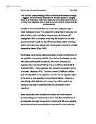
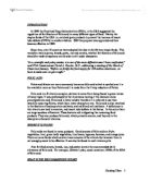
Some types of hybridisation are evolved to be used nowadays in molecular diagnostic assays. The first type is known as Southern Blotting, which was created by Edward Southern1,2,7. Another type of hybridisation technique is known as Northern Blotting. Both techniques are widely used in many areas. Table E shows the characteristics of both types of blotting and how each type of blot is used and figs 2 and 3 below show both types of blotting:
Table E: Characteristics of both northern and southern blots and their respective uses2
Fig 2: Southern blotting2.
First, we electrophorese DNA
fragments in an agarose gel.
Next, we denature the DNA with
base and transfer the single-
stranded DNA fragments from
the gel to a sheet of nitrocellulose
or similar material. We can do this
in two ways: by diffusion, in
which buffer passes through the
gel, carrying the DNA with it
(left), or by electrophoresis (right).
Next, we hybridise the blot to a
labelled probe and detect the
labelled bands by
autoradiography or
phosphorimaging2.
Fig 3: A Northern blot2.
Poly (A)+ RNA was isolated from
The rat tissues indicated on the
right, then equal amounts of
RNA from each tissue were
electrophoresed and Northern
blotted. The RNAs on the blot
were hybridised to a labelled
probe for the rat G3PDH gene,
and the blot was then exposed to
x-ray film. The bands represent
the G3PDH mRNA, and their
intensities are indicative of the
amounts of this mRNA in each tissue.
In overall, nucleic acid hybridisation has helped in many areas as shown in table F below:
Table F: Nucleic acid hybridisation and its uses generally1
In nucleic acid hybridisation, both radioactive (e.g. 32P and 35S) and non-radioactive labelled probes are being used to target nucleic acid1,2,3. However, the total amount of target nucleic acid in a sample is often very low and hence the signal generated by the hybridisation of specific probes is often undetectable1. In order to overcome such limitation, schemes for target amplification (e.g. using PCR) have been developed1. However, no matter how good the schemes are, to produce useful information on the size, identity and quantity of the amplification products, there are some limitations1. One of the main disadvantages is the risk of contaminating untested samples, since the amplification products have to be transferred from the reaction solution to a gel matrix for electrophoresis analysis8. Moreover, the synthesis of non-specific amplification products can be significant when the targets are rare or absent and the sample contains an abundant and diverse nucleic acid hybridisation1. Hence in order to simplify and detect amplified targets in sealed tubes in real time, other types of probes / primers are introduced and listed in table G (fig 4 shows the schematic representation of some nucleic acid hybridisation probes / primers):
Table G: Types of probes / primers, characteristics and their uses1
Fig 4: Schematic representation of fluorescence signal generation with different nucleic acid hybridisation probes1
Fig 4 (continued)1
With these newly introduced probes, nucleic acid hybridisation can be made to detect amplified targets in real time and improves in the techniques of such process.
Overall, nucleic acid hybridisation has been a great help in terms of speed and simplicity1. It can be combined with other molecular biology techniques and can be followed in real time, providing quantitative determination of target nucleic acids over a broad range of concentrations1.
References
1 A.E.M. Salvatore, T. Sanjay & R.K. Fred (2006). Real-time Assays with Molecular Beacons
and Other Fluorescent Nucleic Acid Hybridisation Probes. Clinica Chimica Acta. 363: 48-60.
2 F.W. Robert (2005). Molecular Biology (3rd edition) (International edition). McGraw-Hill co.
3 T.A. Brown (1995). Essential Molecular Biology (A Practical Approach) (Volume 2). Oxford
University Press.
4 L. Ben (2005). Genes VIII. Oxford University Press.
5 B.D. Hall & S. Spiegelman (1961). Sequence Complementarily of T2-DNA and T2-Specific
RNA. Proc. Natl. Acad. Sci. U.S.A. 47: 137-146.
6 A.P. Nygaard & B.D. Hall (1963). A method for the detection of RNA-DNA complexes. J.
Mol. Biol. 9: 125-142.
7 E.M. Southern (1975). Detection of Specific Sequences Among DNA Fragments Separated by
Gel Electrophoresis. Mol. Microbiol. 98: 503-517.
8 S. Kwok & R. Higuchi (1989). Avoiding False Positives with PCR. Nature. 339: 237-238.
9 S. Tyagi & F.R. Kramer (1996). Molecular Beacons: Probes that Fluorescence upon
Hybridisation . Nat. Biotechnol. 14: 303-308.
10 A.P. Nygaard & B.D. Hall (1963). A method for the detection of RNA-DNA complexes. J.
Mol. Biol. 9: 125-142.
11 R.P. Haugland, J. Yguerabide & I. Stryer (1969). Dependence of the Kinetics of Singlet-singlet
Energy Transfer on Special Overlap. Proc. Natl. Acad. Sci. U.S.A. 63: 23-30.
12 C.T. Wittwer, M.G. Herrmann, A.A. Moss & R.P. Rasmussen (1997). Continuous
Fluorescence Monitoring of Rapid Cycle DNA Amplification . BioTechniques. 22: 130-131.
13 A. Tsuji, H. Koshimoto, Y. Sato et al (2000). Direct Observation of Specific Messenger RNA
in a Single Living Cell under a Fluorescence Microscope. Biophys J. 78: 3260-3274.
14 P.M. Holland, R.D. Abratuson, R. Watson & D.H. Glefand (1991). Detection of Specific
Polymerase Chain Reaction Product by Utilizing a 5’-3’ Exonuclease Activity of Thermus
aquaticus DNA Polymerase. Proc. Natl. Acad. Sci. U.S.A. 88: 7276-7280.
15 K.J. Livak, S.J. Flood, J. Mannaro, W. Giusti & K. Dectz (1995). Oligonucleotides with
Fluorescent Dyes at Opposite Ends Provide a Quenched Probe System Useful for Detecting
PCR Product and Nucleic Acid Hybridisation. PCR Methods Appl. 4: 357-362.
16 N. Svanvik, G. Westman, D. Wang & M. Kubista (2000). Lightup Probes: Thiazole Orange-
conjugated Peptide Nucleic Acid for Detection of Target Nucleic Acid in Homogeneous
Solution. Anal Biochem. 281: 26-35.
17 N. Svanvik, A. Stahlberg, U. Schlatedt, R. Sjoback & M. Kubista (2000). Detection of PCR
Products in Real-time using Light-up Probes. Biochem. 287: 179-182.
18 E. Prival, T. Melvin, U. Asseline & P. Vigay (2001). Oligonucleotide-conjugated Thiazole
Orange Probes as “Light-up” Probes for Messenger Ribonucleic Acid Molecules in Living
Cells. Photochem. Photobiol. 74: 532-541.
19 D. Whitcombe, J. Theaker, S.P. Guy,T. Brown & S. Little (1999). Detection of PCR Products
Using Self-probing Amplicons and Fluorescence. Nat. Biotechnol. 17: 804-807.
20 Y. Huang, D. Kong, Y. Yang, R. Niu, H. Shen & H. Mi (2004). Real-time Quantitative Assay
of Telomerase Activity Using the Duplex Scorpion Primer. Biotechnol Lett. 26: 891-895.
21 I.A. Nazarenko, S.K. Bhatnagar & R.J. Hohman (1997). A closed Tube Format for
Amplification and Detection of DNA Based on Energy Transfer. Nucleic Acids Res. 25: 2516-
2521.
22 J. Pickering, A. Bamford, V. Godbole et al (2002). Integration of DNA Ligation and Rolling
Circle Amplification for the Homogeneous, End-point Detection of Single Nucleotide
Polymorphisms. Nucleic Acids Res. 30: E60.
23 J.G. Nadeau, J.B. Pitner, C.P. Linn, J.L. Schram, C.H. Dean & C.M. Nyez (1999). Real-time,
Sequence-specific Detection of Nucleic Acids During Strand Displacement Amplification.
Anal. Biochem. 276: 177-187.
24 D.J. French, C.L. Archard, T. Brown, & D.G. McDowell (2001). HyBeacon Probes: A New
Tool for DNA Sequence Detection and Allele Discrimination. Mol. Cell. Probes. 15: 363-374.
25 Q. Li, G. Luan, Q. Guo & J. Liang (2002). A New Class of Homogeneous Nucleic Acid Probes
Based on Specific Displacement Hybridisation. Nucleic Acids Res. 30: E5.
26 E.R. Kandimalla & S. Agrawal (2000). “Cyclicons” as Hybridisation-based Fluorescent
Primer-probe Synthesis, Properties and Application in Real-time PCR. Bioorg. Med. Chem. 8:
1911-1916.