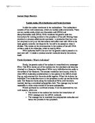
When a sugar bonds together with a nitrogen base, it creates a structure known as a nucleoside. There are five nitrogen bases found in RNA and DNA. These bases are divided into two categories based on their molecular structure
- Purines (adenine & guanine)
- Pyrimidines (thymine, cytosine & uracil)
Diagram 2 shows the nitrogen bases found in RNA and DNA
HOW DNA AND RNA DIFFER
WASTON-CRICK MODEL
In 1953, James Watson and Francis Crick proposed a structure for DNA that:
“ Accounts for this paring of bases but also explains how relatively simply the system of storing and transferring genetic information is.”
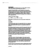
According to Watson and Crick, the shape of a DNA molecule is like a ladder, twisted and coiled into a double helix. The rungs on this ladder would be the nitrogen bases bonded to each other. Adenine always bonds with thymine and is held together by three hydrogen bonds. Cytosine always bonds with guanine and is held together by two hydrogen bonds.
base pairs in DNA
The nitrogen base pairs are bonded to a sugar-phosphate backbone-a chain of alternating sugars and phosphate groups. A nitrogen from the nitrogen bases forms a covalent bond with the first carbon on the sugar molecule which in this case is deoxyribose. The fifth carbon on the sugar bonds with an oxygen from a phosphate group/ Another oxygen from the same phosphate group bonds with carbon-3 on the sugar molecule, and it is this repeating chain that makes up the backbone of a DNA strand. But since DNA is double stranded there are two sugar phosphate backbone in DNA molecule.
DNA REPLICATION
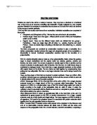
DNA replication begins with a partial unwinding of the double helix at an area known as the replication fork. An enzyme known as DNA helicase accomplishes this unwinding. As the two DNA strands separate and the bases are exposed the enzyme, DNA ploymerase moves in at the point of synthesis will begin.
The DNA polymerase knows where to start the synthesis due to a short segment of RNA known as RNA primer. The primer is “laid down” complementary to the DNA template by an enzyme known as RNA polymersase.
The DNA polymerase then adds nucleotides one by one in an exactly complementary manner: A to T and C to G.
Diagram to show DNA replication
DNA polymerase is described as being “template dependent” in that it will read the sequence of the bases on the template strand and then synthesize the strand. DNA polymerase catalyzes the formation of the hydrogen bonds between each arriving nucleotide and the nucleotides on the template strand.
STRUCTURE AND FUNCTION OF RNA
RNA is structurally similar to DNA but it differs in function. DNA has only one function-storing genetic information in its sequence of nucleotide bases. But there are three main kinds of ribonucleic acid (RNA), each of which carries out a specific job.
RIBOSOMAL RNA- exists outside the nucleus in the cytoplasm of the cell in structures called ribosomes. Ribosome is where protein synthesis takes place.
MESSENGER RNA- is the nucleic acids that “record” information from DNA in the cell nucleus and carry it to the ribosome.
TRANSFER RNA- the function of transfer RNA is to deliver amino acids one by one to protein chains growing at the ribosome.
RNA SYNTHESIS: TRANSCRPTION
The process of converting information contained in a DNA segment into proteins begins with the synthesis of messenger RNA molecules containing anything from several hundred to several thousand ribonucleotides, depending on the size of the protein.
“Each of the 100,000 or so protein in the human body is synthesized from a different messenger RNA that has been transcribed from a specific gene on the DNA”
In eukaryotic cells DNA is very important and if damaged would change or mutate the coding sequence. This could greatly affect the cell or even the whole organism.
If DNA were to venture out into the cytoplasm it would be more vulnerable to damage from:
- Chemicals
- Ultra violet light
- Other agents
Therefore, messenger RNA does the venturing for DNA.
Messenger RNA is synthesized in the cell nucleus by transcription of DNA, a similar process to DNA replication. As in DNA replication a small section of the DNA helix unwinds, and the bases on the two strands are exposed. RNA nucleotides line up in the proper order, by hydrogen bonding, to their complementary bases on the DNA. The nucleotides are joined together by a DNA dependent RNA polymerase enzyme. As a result, messenger RNA is formed.
But how does the enzyme know where one gene start and stop and another begin. How does the enzyme know where to start? The starting point of a gene is marked by a certain base sequence called a promoter site. These sites are recognized by a factor known as sigma. It is sigma’s job to recognize the promoter sites and “tell” the DNA dependent RNA polymerase where to begin the process of transcription.
There is also a base sequence at the end of a gene that stops messenger RNA synthesis. A factor that helps signal the end of a gene is called rho. When the end of the gene is near, the rho factor binds to the messenger RNA and interacts with the RNA polymerase. This causes the enyme to denture and fall off, thus stopping transcription.
PROTEIN SYNTHESIS
Most of the time when a cell is not dividing, it is performing a series of activities under the control of the DNA, in the nucleus. In order to do this, information from certain portions of the DNA in the chromosomes must be taken out into the cytoplasm, to be used to make (synthesize) control proteins (enzymes ect.) for the cell. This process is known as protein synthesis.
STEP 1 OF PROTEIN SYNTHESIS: INTIATION
Protein synthesis is intitated when an messenger RNA, a ribosome and the first transfer RNA molecule carry its Methionine amino acid, come together.