Intracellular Ca2+ also causes the expression of CD62 (P-selectin) to form aggregates of platelets. These aggregates, again by Ca2+ signalling, express outer membrane phosphatidylserine, a classical apoptotic signal. In this circumstance it is also a coagulation signal, as it creates an unusually strong negative outer membranous charge, attracting coagulation factors such as factors Va and Xa to form thrombin from prothrombin. This thrombin formation through the aggregate further increases the activation already discussed, but also cleaves fibrinogen to form fibrin. This stabilises the aggregate to the status of a clot, which forms a plug once it retracts. During the course of this, microparticles (40nm x 1µm) are also released as exocytotic buds from the platelets. These are part of the apoptotic process, but are capable of the functions outlined above, and thus contribute to the role of platelets in haemostasis.[3,6,7]
There are a few methods by which these crucial particles are obtained from donors to be prepared for transfusion into cancer or thrombocytopenic patients. The standard whole blood derived platelet rich plasma (WBD-PRP) comes from normal donations. A closed system donation of the 450ml unit is received, and is soft spun producing a platelet concentrate in the supernatant. This supernatant is then hard spun to produce a platelet sediment, and approximately 4-8 of these are combined to give 1 platelet unit. This is a comparatively cheaper method to use, but exposes the recipient to an increased number of donors, thus presenting more potential for transfusion reactions (TRs) or infections. A Buffy coat can be used, which uses the same method as WBD-PRP but exchanges the spin order i.e. hard spin first, soft spin second, and finally apheresis can be used. Apheresis is the most expensive procedure, and reduces the amount of blood components received, this also reduces the number of donors as well as the risk of TRs or infections.[1,8] It is important to have leukoreduced units of platelets as nearly 90% of TRs are cytokine induced, and 10% by antibodies. Thus, the TRAP Study Group strongly encourage the treatment of units with UVB light, or amotosalen, to reduce the number of active leukocytes present.[9,10]
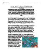
Ratio of WBD donors vs.Apheresis donors with
respect to platelets recovered [17]
In addition to this, platelet function must be maintained without contamination, so the polyolefin bags used (such as PO-80), must have a large surface area, be thin and of an adequate material for O2/CO2 exchange to promote aerobic metabolism and ATP maintenance. A drop in ATP concentration by 30%, or to below 4µmol/1011 PLT can be deemed notably detrimental to function and morphology and this can be detected by pH testing.[9] Anaerobic respiration will cause a rapid drop in glucose and a rise in lactate which displaces bicarbonate, thus reducing the pH. So a range of pH6.2-7.4 has been generally attributed as satisfactory.[1,8,9,11,12,13] To enable good O2 access, and avoid sedimentation, oscillating agitation must be maintained at 22oC,[12] and due to potential bacterial survival and growth at this temperature, a shelf life of 5 days for non-bacterial culture tested samples has been set.[1,3,4,6,14]
Platelets are made in the body, and are designed to resist any insult to the body’s integrity, however the removal of anything from the body itself is an insult. So from the point of donation platelets experience some degree of activation, and for the past 20 years and still today efforts are made to resist this activation, also known as the platelet storage lesion (PSL). Shrivastava describes the PSL rather exhaustively as “the sum of all deleterious changes leading to progressive damage in platelet structure and function that arise from the time blood is drawn from a donor to the time platelets are transfused to a recipient.”[4] As can be surmised from this statement, the PSL refers to morphological, metabolic and apoptotic changes, all affecting the haemostatic function, the activation status and activation potential of the platelets. This starts and progresses from the point of donation, thus fresher platelets are better for transfusions.[3,4,6,9,11]
Generally, the PSL consists of morphological changes and a reduction in the platelets’ physiological responsiveness to typical agonists. The most commonly utilised change is the morphological change from smooth discoid platelets, to spiny spheres potentially extending pseudopodia and releasing microparticles. This is due to actin and microtubule remodelling, caused by Ca2+ release from the DTS by mechanisms previously described, and thus highlights the activated status in the PSL. This can be affected by the type of collection, with apheresis promoting less of a PSL than WBD-PRP or Buffy coats, due to the amount of shear stress imposed on the platelets.[3,4,9] Following collection, oscillating agitation should be adopted, as this reduces the amount of foreign surface contact the platelets experience with the container, thus reducing their activation.[12] The fact that the platelets are activated reduces their potential for further activation in the recipient, thus the PSL is characterised by a rise in activation, and a reduction in future activation potential.
Not a vast amount is known about the PSL at a biochemical level but a few major aspects have been elucidated. A seemingly common occurrence in the PSL is an alteration to the vWF binding complex (GPIb/GPV/GPIX). This reduces the platelets ability to adhere to vWF, and thus the potential for further activation and aggregation. This could be due to metalloproteinase release by the platelet in response to shear stress, causing GPIbα cleavage. The complex also commonly clusters, creating a ligand for the phagocytosis-inducing αMβ2 receptor on macrophages. This provides an additional ‘apoptotic’ signal for the recipient’s Küppfer cells to use to target the platelets, along with the CD62 and the elevated phosphatidylserine, to remove them from the circulation. This is particularly heightened in cold storage, e.g. at 4oC, leading to 27% reduction in platelet recovery and circulatory survival after 72 hours storage, highlighting why this is not a viable storage condition.[4] The platelets are also characteristically desensitised to agonistic stimulation. This can be caused by P2Y1 and P2Y12, and P2X1 receptor desensitisation to ADP and ATP respectively,[6] and the production of calpalin containing microvesicles. This compound degrades actin, talin, tubulin, and vinculin reducing the platelet’s ability to change morphology, thus removing its response to pro-coagulant agonists.[4]
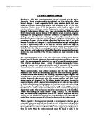
It is important to discount platelets displaying advanced PSL for transfusion. There is no set automated test as yet, but a few standard ones that are employed. There are 2 that are focussed on in a clinical setting, and these are swirling and pH testing. Both are simple to perform, accurate but indirect assessments. Discoid platelets have a non-uniform morphology, thus when they move in agitation, they can scatter or diffract light. PSL platelets have pseudopodia and/or are spherical, which removes this ability. Thus when agitated,
the display of ‘swirling’ provides an indirect
Image of platelet swirling. The scattering of light photons indication of good quality platelets.[1,3,12] If a pH
creates a non uniform presentation of the light passing value outside the range of pH6.2-7.4 is detected,
through the discoid platelets in the sample [18] this is evidence enough of a high lactate
concentration and metabolically unviable platelets.[1,6]
More complex tests can be performed, but these are typically reserved for the research setting. Flow cytometric analysis of microparticle presence, aggregation and surface marker presence such as GPIb or phosphatidylserine (using Annexin V) has been used in some studies,[4,6,15] or light scattering ability. This however assesses uniformity in static light scattering, as in normal flow cytometry, without marker analysis. Dynamic light scattering is much more useful, as instead of the target platelet being in a ‘fixed focus point,’ they move. More fluctuations in light intensity highlights more diverse contours on the membrane, and so a discoid morphology. This applies the same theory as the swirling test.[6]
‘Pre’ and ‘post’ treatment comparisons have been used also, such as treating a sample with an agonist such as ADP, or EDTA (normally an anticoagulant but as it is a Ca2+ chelator it induces apoptotic and pro-coagulation actions[11]) to cause changes such as in mean platelet volume (MPV). A major difference between non-EDTA MPV and EDTA-treated MPV reveals high activation potential, and thus transfusion viability.[6,7,11,12,15] Hypotonic shock response has also been used in this way.[6,9] These all focus on the PSL, however the shelf life of platelet is determined by the potential of bacterial contamination. Testing for bacteria could potentially extend the limit from 5 days to 7 or 9 days, if a negative result is presented. As some of the more common infections, such as S. epidermidis, have slow incubation times, a later test would be required, but a conclusive test at day 4 would reveal 97.9% potential infections. So, if a slow, yet accurate test was started at day 1 or 2 to be concluded at day 3 or 4, shelf life could be justifiably extended, making the PSL the length determining factor, not the possibility of bacterial contamination.[14]
Should the above proposal of early bacterial and viral testing be producible, the focus then would be the inhibition or prolonging of the PSL. Addition of inhibitors such as prostacyclin to stop ADP release, cytochalasin B to prevent actin remodelling, or metalloproteinase inhibitors to prevent the vWF receptor complexing are viable suggestions, however the issue of their removal, and platelet reactivation needs considered.[4,6] Metabolic arrest induced by anti-mycin A for 1 hour, then 4oC storage for 48 hours, followed by warming in a high glucose concentration medium has been proposed to correct this, but this highlights the requirement of a high level of familiarity and expertise required by technicians[6].
Platelets are invaluable in medicine, particularly for those undergoing treatment for cancers, and thrombocytopenia. However, their high value is equated with a high level of care and treatment to guarantee their integrity and viability for those patients who need them. The above suggestions reveal the requirement for novel, innovative, drastic techniques to help maintain the limited stock of platelets transfusion services possess for as long as possible, retaining sterility from microbes, including viruses, and their activation potential.
Bibliography
[1] STRONCEK, D.F. and REBULLA, P., 2007. Platelet transfusions. Lancet, 370(9585), 427-438
[2] PAVENSKI, K., WARKENTIN, T.E., SHEN, H., LIU, Y. and HEDDLE, N.M., 2010. Posttransfusion platelet count increments after ABO-compatible versus ABO-incompatible platelet transfusions in noncancer patients: an observational study. Transfusion,
[3] MAURER-SPUREJ, E. and CHIPPERFIELD, K., 2007. Past and future approaches to assess the quality of platelets for transfusion. Transfusion medicine reviews, 21(4), 295-306
[4] SHRIVASTAVA, M., 2009. The platelet storage lesion. Transfusion and apheresis science : official journal of the World Apheresis Association : official journal of the European Society for Haemapheresis, 41(2), 105-113
[5] HERR, A.B. and FARNDALE, R.W., 2009. Structural insights into the interactions between platelet receptors and fibrillar collagen. The Journal of biological chemistry, 284(30), 19781-19785
[6] CAUWENBERGHS, S., VAN PAMPUS, E., CURVERS, J., AKKERMAN, J.W. and HEEMSKERK, J.W., 2007. Hemostatic and signaling functions of transfused platelets. Transfusion medicine reviews, 21(4), 287-294
[7] WEI, A.H., SCHOENWAELDER, S.M., ANDREWS, R.K. and JACKSON, S.P., 2009. New insights into the haemostatic function of platelets. British journal of haematology, 147(4), 415-430
[8] HEDDLE, N.M., ARNOLD, D.M., BOYE, D., WEBERT, K.E., RESZ, I. and DUMONT, L.J., 2008. Comparing the efficacy and safety of apheresis and whole blood-derived platelet transfusions: a systematic review. Transfusion, 48(7), 1447-1458
[9] SHANWELL, A., DIEDRICH, B., FALKER, C., JANSSON, B., SANDGREN, P., SUNDKVIST, L., SVENSSON, L., VESTERINEN, M. and GULLIKSSON, H., 2006. Paired in vitro and in vivo comparison of apheresis platelet concentrates stored in platelet additive solution for 1 versus 7 days. Transfusion, 46(6), 973-979
[10] Leukocyte reduction and ultraviolet B irradiation of platelets to prevent alloimmunization and refractoriness to platelet transfusions. The Trial to Reduce Alloimmunization to Platelets Study Group. 1997. The New England journal of medicine, 337(26), 1861-1869
[11] SINGH, H., CHAUDHARY, R. and RAY, V., 2003. Evaluation of platelet storage lesions in platelet concentrates stored for seven days. The Indian journal of medical research, 118, 243-246
[12] HOLME, S., VAIDJA, K. and MURPHY, S., 1978. Platelet storage at 22 degrees C: effect of type of agitation on morphology, viability, and function in vitro. Blood, 52(2), 425-435
[13] EZUKI, S., KANNO, T., OHTO, H., HERSCHEL, L., ITO, T., KAWABATA, K., SEINO, O., IKEDA, K. and NOLLET, K.E., 2008. Survival and recovery of apheresis platelets stored in a polyolefin container with high oxygen permeability. Vox sanguinis, 94(4), 292-298
[14] BRECHER, M.E., HOLLAND, P.V., PINEDA, A.A., TEGTMEIER, G.E. and YOMTOVIAN, R., 2000. Growth of bacteria in inoculated platelets: implications for bacteria detection and the extension of platelet storage. Transfusion, 40(11), 1308-1312
[15] SEGHATCHIAN, J., 2006. A new platelet storage lesion index based on paired samples, without and with EDTA and cell counting: comparison of three types of leukoreduced preparations. Transfusion and apheresis science : official journal of the World Apheresis Association : official journal of the European Society for Haemapheresis, 35(3), 283-292
[16] Accessed: 21:14, 09/04/2010
[17] Accessed: 21:07, 09/04/2010
[18] Accessed: 21:24, 09/04/2010