In the Golgi apparatus, cysteines proximal to the C-terminal methyl-isoprenyl-cysteine are palmitoylated, by the receipt of a 14 carbon fatty acid called palmitate. This can be added to 1 or more cysteines by thioester bonds, but typically 2 cysteine residues receive this PTM. The now fully modified GTPase is translocated to the corresponding membrane, usually the PM, via the biosynthetic-secretory pathway, to be incorporated, mostly, into lipid rafts.3 These palmitate moieties enable cytosolic anchorage of the Ras proteins into the PM, however K-Ras proteins do not require such moieties, as they contain positively charged polybasic tracts, typically of lysine, which can electrostatically associate with the negative phosphate heads of the PM. Due to the nature of the association, there is a low half-time for K-Ras association with the PM, typically in minutes. It is relatively easily removed, by strategic phosphorylation by PKC, disrupting these balanced interactions, and it is then sequestered into mitochondria, the Golgi apparatus or ER.3, 6, 13
Not only are these PTMs required for activity, they can also define the GTPase’s specific function e.g. if RhoB receives a geranylgeranyl group it is directed to endomembranes, but is sent to the PM if possessing a farnesyl group. Also, subtle differences in the polybasic tract lead to different localisations too, as Rac1 is directed to lipid rafts, Rac2 to endosomes and RhoG to calveolar vesicles.13 Rap1 highlights the importance of spatiotemporal relationships for GTPases, guanine nucleotide exchange factors (GEFs) and GTPase activating proteins (GAPs), as it is localised not at the PM, but on cellular Ca2+ stores such as mitochondria. This is directed by a phosphorylated serine at its C-terminal. Ca2+ can be released at specific points in time and subcellular locations, and Rap1 is activated by second messengers such as Ca2+, and this promotes a fast response. Also other Rap proteins used in thromboxane A2, and thrombin responses are localised proximal to RTKs on the PM.3, 5
Despite the transient nature of Ras signal transduction, the GTPase activity of Ras is not rapid enough to convert the Ras-GTP to inactive Ras-GDP at a rate suitable for the cell. However, this therefore allows for manipulation of Ras’ GTPase activity, and this is by GEFs, and GAPs. GEFs are corporated into Ras signalling complexes, as Ras activity activates them, creating a positive feedback loop. They facilitate the removal of GDP from the G domain, and due to the high cellular concentration of GTP, of ~1mM, this is rapidly replaced by GTP, activating the Ras protein’s signalling capabilities.11 Conversely, GAPs possess a specific Arg or Gln finger which, when inserted into the GTPase, raises the GTPase activity significantly,11 and GAPs also prevent effecters binding to the Ras, as they blocks the docking sites and switches. Thus, GEFs favour activation of Ras signalling, and GAPs favour inactivation of Ras signalling.3-5, 7, 10
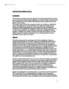
These GEFs and GAPs target specific Ras proteins, and this is shown by the fact that the Rho subfamily has over 60 GEFs and 70 GAPs for their 70 members.10, 12 There has to be very tight control over Ras due to its pro-oncogenic nature. 30% of human tumours have some form of mutation that heightens Ras activity and expression.5, 7, 8 This requires that GEFs and GAPs are controlled, and this is typically by Ca2+ and diacylglycerol (DAG), and so by phospholipase C (PLC) further upstream, and especially PLCε due to its dual GEF and PLC activities.4, 14 DAG activates PKC and some GEFs e.g. RasGRF1-CDC25, but also the release of calcium (due to IP3 complexing with its receptor on Ca2+ stores) will cause translocation of GAPs, particularly CAPRI and RASAL which are sensitive to Ca2+ amplitude and oscillations respectively. They possess active N-terminal C2 domains, requiring Ca2+ before they can anchor to the PM.8 The best known pathway is the growth MEK/Erk MAPK pathway induced by Ras following EGF signalling. It is also used by Rac, and Rac’s activity is modulated by calmodulin under Ca2+ control. A Ras GEF called CalDAG-GEF1 is activated by
Ras-MAPK Pathway 20
DAG and Ca2+, through the use of calmodulin. This was first identified in T cells, and overexpression of this GEF leads to very aggressive leukaemia.4
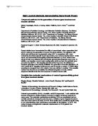
The Ras superfamily coordinates many cellular functions, with subfamilies within the Ras family each controlling specific aspects of cellular activity. There are 10 subfamilies,15 but the main 5 are Ras Sarcoma (Ras) which control differentiation, proliferation and survival; Ras Homology (Rho) responsible for actin organisation, cell migration, cell cycle progression and morphology; Ras-like proteins in brain (Rab) control vesicular trafficking from vesicle formation to fusion with target compartments; Ras-like nuclear proteins (Ran) enable nuclear import and export using importin and exportin respectively. It is the most common Ras protein, having only 1 type in the subfamily; and ADP-ribosylation-factor (Arf) which possesses an additional N-terminal region, compared to other Ras proteins, for myristate group interaction for anchorage, and aids vesicle transport complex formation e.g. COPI-Arf1.10, 12, 15
Subfamilies within the Ras Superfamily 21
Focussing on the Rho family, these cause cytoskeletal changes, facilitating changes in morphology and migration. The family was identified in 1985, and since then, members Rho, Rac and CDC42 have been well characterised, being responsible for stress fibre formation, lamellipodia formation and filopodia formation respectively, all using separate signalling pathways. Despite the use of separate pathways, there is crosstalk and co-operation between the GTPases, for example whilst Rac may induce actin polymerisation and destabilisation of microtubules, CDC42 alters the location of the microtubule organisation centre to compensate for the movement of the cell, stabilises Rac and also interprets chemotactic signals. So, CDC42 in effect defines the direction of movement, enables movement, and controls subcellular compartmental localisation whilst migrating, and Rac itself causes the movement. In the cell cycle, Rho, Rac and CDC42 are all used for progression, and strongly highlight their co-dependence, not for activity but for the ability to use this activity. As Rho inhibits p21 expression it also causes Cyclin D1 transcription. Rac then enables Cyclin D1 expression in G1 by promoting the translation of the resultant mRNA. This shows the reliance of Rac on the activity of Rho, for the lack of 1 GTPase would cause the cascade to collapse. Finally, in phagocytosis, Rho is used for the pseudopodia formation in both type I and II, but Rac is only used in type I. This is because type I phagocytosis induces an inflammatory response to pathogens, and Rac is part of the phagosome NADPH oxidase complex, whereas type II only uses Rho as part of the apoptosis ‘clean up.’ Employment of Rac GTPases, even within subfamilies, is therefore very specific.12, 16, 17, 18
Rab proteins are the largest subfamily of Ras, having over 60 members, targeting distinct membranes for vesicular trafficking. They recruit effecters to sites of vesicular budding e.g. Rab9 recruits TIP47 to mannose-6-phosphate receptors to target for retrograde recycling to the Trans Golgi Network. Prior to fusion of vesicles to targets, they associate with SNARE proteins either directly e.g. Rab5 is an intermediate molecule, connecting a SNARE and Sec1, or facilitate the direct interaction of the 2 SNAREs communities, and then cause uncoating. This is to enable the proximity required to expel the water molecules from the fusion site. Rab5 recruits GAPVD1 to clathrin complexes, which dephosphorylates the µ2 subunit of the AP2 coat, destabilising it to dissociate. Different Rab proteins are localised to different organelles, and microdomains on the organelles. Intrinsic GEFs in these domains enrich these, and Guanine nucleotide dissociation inhibitors, GDIs (prevent GDP release), cause sequestering of the Rab proteins, promoting cycling. The main microdomains contain Rab5, Rab4 and Rab5, or Rab4 and Rab11, and this could be caused by coupling as 1 effecter may bind or need several Rab variants. This also applies to other families, as Ras and Rap1 are known to share effecters, but in that case, the signalling pathways used and end phenotypic responses are not the same.3 As Rabs recruit phospholipid, and phosphoinositol kinases and phosphatases, they also affect membrane compositions and identity.10, 11, 19
Despite the homology of the proteins in the Ras superfamily that validates them as members, there are key differences in structure and specificity between the subfamilies and the group members, making them non-redundant, specific modulators of cell activity. They require acute control to avoid disease states, most notably cancers, but they also are required for a cell to fully survive and function. It can be seen by studying the areas of control individual subfamilies have, and even that individual Ras GTPases have, that the loss of 1 controllable, functioning protein, through genetic mutation or another method of disruption, can cause the cell to die, become non-functional, or hyperactivited in a specific realm. This potentially could lead to catastrophe, almost certainly for the surrounding cellular population, but the organism as a whole. Due to the strong prevalence of Ras mutations or dysfunctions in human cancers, research into novel treatments may need to divert its focus from signal induction, to Ras transduction.
References
1. Ellis RW et al. The p21 src genes of Harvey and Kirsten murine sarcoma viruses originate from
divergent members of a family of normal vertebrate genes. Nature 1981; 292: 506–511.
2. Shimizu K, Goldfarb M, Perucho M & Wigler M. Isolation and preliminary characterization of the
transforming gene of a human neuroblastoma cell line. roc. Natl Acad. Sci. USA 1983; 80: 383– 387.
3. Zwartkruis FJ, Bos JL. Ras and Rap1: Two highly related small GTPases with distinct function. Exp
Cell Res 1999 Nov 25;253(1):157-65.
4. Buday L, Downward J. Many faces of ras activation. Biochim Biophys Acta 2008 Dec;1786(2):178-87.
5. Walker SA, Cullen PJ, Taylor JA, Lockyer PJ. Control of ras cycling by Ca2+. FEBS Lett 2003 Jul 3;546(1):6-10.
6. Rocks O, Peyker A, Bastiaens PI. Spatio-temporal segregation of ras signals: One ship, three anchors, many harbors. Curr Opin Cell Biol 2006 Aug;18(4):351-7.
7. Goldfinger LE. Choose your own path: Specificity in ras GTPase signaling. Mol Biosyst 2008 Apr;4(4):293-9.
8. Yarwood S, Bouyoucef-Cherchalli D, Cullen PJ, Kupzig S. The GAP1 family of GTPase-activating proteins: Spatial and temporal regulators of small GTPase signalling. Biochem Soc Trans 2006 Nov;34(Pt 5):846-50.
9. Vetter IR, Wittinghofer A. The guanine nucleotide-binding switch in three dimensions. Science 2001 Nov 9;294(5545):1299-304.
10. Wennerberg K, Rossman KL, Der CJ. The ras superfamily at a glance. J Cell Sci 2005 Mar 1;118(Pt 5):843-6.
11. Stenmark H. Rab GTPases as coordinators of vesicle traffic. Nat Rev Mol Cell Biol 2009 Aug;10(8):513-25.
12. Etienne-Manneville S, Hall A. Rho GTPases in cell biology. Nature 2002 Dec 12;420(6916):629-35.
13. Bustelo XR, Sauzeau V, Berenjeno IM. GTP-binding proteins of the Rho/Rac family: Regulation, effectors and functions in vivo. Bioessays 2007 Apr;29(4):356-70.
14. Bunney TD, Baxendale RW, Katan M. Regulatory links between PLC enzymes and ras superfamily GTPases: Signalling via PLCepsilon. Adv Enzyme Regul 2009;49(1):54-8.
15. Munemitsu S, Innis M, Clark R, McCormick F, Ullrich A, Polakis P. (1990). Molecular cloning and
experssion of a G25K cDNA, the human homolog of the yeast cell cycle gene CDC42. Mol Cell Biol 1990; 10 (11): 5977–82.
16. Caron E, Hall A. Identification of two distinct mechanisms of phagocytosis controlled by different rho GTPases. Science 1998 Nov 27;282(5394):1717-21.
17. Ng J, Nardine T, Harms M, Tzu J, Goldstein A, Sun Y, Dietzl G, Dickson BJ, Luo L. Rac GTPases control axon growth, guidance and branching. Nature 2002 Mar 28;416(6879):442-7.
18. Sugihara K, Asano S, Tanaka K, Iwamatsu A, Okawa K, Ohta Y. The exocyst complex binds the small GTPase RalA to mediate filopodia formation. Nat Cell Biol 2002 Jan;4(1):73-8.
19. Caswell PT, Spence HJ, Parsons M, White DP, Clark K, Cheng KW, Mills GB, Humphries MJ, Messent AJ, Anderson KI, McCaffrey MW, Ozanne BW, Norman JC. Rab25 associates with α5β1 integrin to promote invasive migration in 3D microenvironments. Dev Cel 2007 Oct; 13: 496-510
20. Accessed: 23:43, 23/04/2010
21. Accessed: 23:54, 23/04/2010