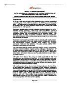
Using stains on expectorate samples from (also called sputum), scientists can identify MTB under a regular microscope. Since MTB retains certain stains after being treated with acidic solution, it is classified as an (AFB). The most common acid-fast staining technique, the , dyes AFBs a bright red that stands out clearly against a blue background. Other ways to visualize AFBs include an and .[1][7][8]
1.4 Transmission
When people suffering from active pulmonary TB cough, sneeze, speak, or spit, they expel infectious droplets 0.5 to 5 in diameter. A single sneeze can release up to 40,000 droplets. Each one of these droplets may transmit the disease, since the infectious dose of tuberculosis is very low and the inhalation of just a single bacterium can cause a new infection.[6]
People with prolonged, frequent, or intense contact are at particularly high risk of becoming infected, with an estimated 22% infection rate. A person with active but untreated tuberculosis can infect 10–15 other people per year. Others at risk include people in areas where TB is common, people who inject drugs using unsanitary needles, residents and employees of high-risk congregate settings, medically under-served and low-income populations, high-risk racial or ethnic minority populations, children exposed to adults in high-risk categories, patients by conditions such as /, people who take immunosuppressant drugs, and health care workers serving these high-risk clients.[7]
Transmission can only occur from people with active — not latent — TB. The probability of transmission from one person to another depends upon the number of infectious droplets expelled by a carrier, the effectiveness of ventilation, the duration of exposure, and the of the M. tuberculosis . The chain of transmission can, therefore, be broken by isolating patients with active disease and starting effective anti-tuberculous therapy. After two weeks of such treatment, people with active TB generally cease to be contagious. If someone does become infected, then it will take at least 21 days, or three to four weeks, before the newly infected person can transmit the disease to others. TB can also be transmitted by eating meat infected with TB. Mycobacterium bovis causes TB in cattle. [3][8][9]
-
Primary Tuberculosis:
Primary Tuberculosis is an infection of persons who have not had prior contact with the tubercle bacillus. The bacilli usually enter the body by inhalation but can also enter through the gastrointestinal tract or by cutaneous or subcutaneous inoculation.
Inhaled bacilli are commonly deposited in alveoli immediately beneath the pleura, usually in the lower part of the upper lobes or the upper part of the lower lobes. These areas receive the greatest volume of inspired air. The initial infection produces only slight abnormalities and may cause only slight malaise and mild fever.[1][7]
Since sensitized T cells are lacking, the tubercle bacilli multiply freely and enter the blood stream and lymphatics. Cell-mediated immunity develops over a period of 3 to 6 weeks.
The primary infection characteristically produces a “Ghon complex”- that is, a single lesion in the pulmonary parenchyma, usually subpleural, that is accompanied by a lesion of the hilar lymph nodes draining that part of the lung. The primary lesion, or Ghon focus, in the lung is typically a 1cm, grayish, circumscribed nodule. It becomes granulomatous within a few days and by the second week has soft caseous, necrotic center.[3]
Tubercle bacilli drain through lymphatic channels to infect the hilar lymph nodes and form the second part of the Ghon complex. In over 90% of normal adults the infection follows this self-limited course, because the cellular immune response is sufficient to control the multiplication of bacilli.[3]
Therefore, in both the lung and the lymph nodes the lesions of the Ghon complex heal, undergoing shrinkage, fibrous scarring, and calcification.
Most of the organisms die, but a few remain viable for years. Later, if immune mechanisms wane or fail, the resting bacilli may break out and cause serious tuberculous infection.
Progressive primary tuberculosis is a rarer alternative course, in which the immune response fails to control multiplication of the tubercle bacilli.Infection takes this course in less than 10% of normal adults, but it is common in children under 5 years of age.
In adults, progressive primary tuberculosis most commonly occurs in patients with suppressed or defective immunity.
The primary Ghon focus in the lung enlarges rapidly, erodes the bronchial tree, and spreads, a sequence that results in adjacent “satellite” lesions.
This process is accompanied by caseous enlargement of the hilar lymph nodes, which may erode through the wall of the bronchus and discharge bacilli, thereby producing tuberculous pneumonia.
Clinical manifestations are abrupt high fever (associated with progression to tuberculous pneumonia), pleurisy with effusion, and lymphadenitis. The highly active lesions may seed the blood stream with tubercle bacilli and result in life-threatening dissemination of the bacilli.[1][8][9]
- Secondary Tuberculosis:
Secondary (cavitary) tuberculosis usually results from reactivation of dormant, endogenous tubercle bacilli in a sensitized patient who has had previous contact with the tubercle bacillus. In some cases, the disease is caused by reinfection with exogenous bacilli.
Secondary tuberculosis may develop any time after primary infection, even decades later. Reactivation typically begins in the apical or posterior segments of one or both upper lobes (Simon’s foci), where the organisms were seeded during the primary infection.
Radiographically, the lesions are spherical and cavitary- the so-called coin lesions. A fibrous capsule surrounds the caseous, acellular center, which contains numerous tubercle bacilli. From these cavitary nodules the organisms can spread through the lung and be discharged into the air during bouts of coughing.[8][9]
The symptoms of secondary tuberculosis begin with cough, which may be erroneously attributed to smoking or to a “cold”. Low-grade fever develops, with general malaise, fatigue, anorexia, weight loss, and often night sweats.
As the disease progresses, the cough worsens and the sputum may be streaked with blood. The rupture of a branch of the pulmonary artery in the wall of a cavity leads to massive hemoptysis and asphyxiation or exanguination.[13][14]
These pulmonary lesions of secondary tuberculosis are often complicated by a variety of secondary effects, including :
(1) Scarring and calcification
(2) Spread to other areas
(3) Pleural fibrosis and adhesions, with associated pleurisy, sharp pleuritic pain, and shortness of breath
(4) Rupture of a caseous lesion, which spills bacilli into the pleural cavity
(5) Erosion into a bronchus, which seeds the mucosal lining of bronchioles, bronchi, and trachea
(6) Implantation of bacilli in the larynx, which causes laryngitis, hoarseness, and pain on swallowing[12]
Lesions of secondary tuberculosis acquired through the gastrointestinal tract (usually with M. t. bovis) can lead to entrapment of bacilli in lymphoid patches of small and large bowel
- Pathology of Pulmonary Tuberculosis
4.1 Main symptoms of pulmonary tuberculosis
When the disease becomes active, 75% of the cases are TB. Symptoms include chest pain, , and a productive, prolonged cough for more than three weeks. Systemic symptoms include fever, chills, , , weight loss, pallor, and often a tendency to fatigue very easily. [6]
4.2 General features
Mycobacterium tuberculosis is the organism that is the causative agent for tuberculosis (TB). There are other "atypical" mycobacteria such as M. kansasii that may produced a similar clincal and pathologic appearance of disease. M. avium-intracellulare (MAI) seen in immunocompromised hosts (particularly in persons with AIDS) is not primarily a pulmonary infection in terms of its organ distribution (mostly in organs of the mononuclear phagocyte system).[6][8]
4.3 Patterns of Infection
There are two major patterns of disease with TB which are primary tuberculosis and secondary tuberculosis.
Primary tuberculosis: seen as an initial infection, usually in children. The initial focus of infection is a small subpleural granuloma accompanied by granulomatous hilar lymph node infection. Together, these make up the Ghon complex. In nearly all cases, these granulomas resolve and there is no further spread of the infection.
Secondary tuberculosis: seen mostly in adults as a reactivation of previous infection (or reinfection), particularly when health status declines. The granulomatous inflammation is much more florid and widespread. Typically, the upper lung lobes are most affected, and cavitation can occur.
When resistance to infection is particularly poor, a "miliary" pattern of spread can occur in which there are a myriad of small millet seed (1-3 mm) sized granulomas, either in lung or in other organs.
The following illustrate gross pathologic findings with tuberculosis:
4.4 Microscopic Findings
Microscopically, the inflammation produced with TB infection is granulomatous, with epithelioid macrophages and Langhans giant cells along with lymphocytes, plasma cells, maybe a few PMN's, fibroblasts with collagen, and characteristic caseous necrosis in the center. The inflammatory response is mediated by a type IV hypersensitivity reaction. This can be utilized as a basis for diagnosis by a TB skin test. An acid fast stain (Ziehl-Neelsen or Kinyoun's acid fast stains) will show the organisms as slender red rods. An auramine stain of the organisms as viewed under fluorescence microscopy will be easier to screen and more organisms will be apparent. The most common specimen screened is sputum, but the histologic stains can also be performed on tissues or other body fluids. Culture of sputum or tissues or other body fluids can be done to determine drug sensitivities.[13]
4.5 Physical
Primary tuberculosis is characterized by the absence of any signs on clinical evaluation. These patients are identified by a positive TST result. Tuberculin hypersensitivity may be associated with erythema nodosum and phlyctenular conjunctivitis.
Signs of disease depend on the site involved (pulmonary or extrapulmonary).
Pulmonary disease may manifest itself in several forms, including endobronchial tuberculosis with focal lymphadenopathy, progressive pulmonary disease, pleural involvement, and reactivated pulmonary disease.
Endobronchial disease: Enlargement of lymph nodes may result in signs suggestive of bronchial obstruction or hemidiaphragmatic paralysis. Vocal cord paralysis may occur as a result of local nerve compression. Dysphagia due to esophageal compression also may be observed.[12][14]
Progressive primary pulmonary tuberculosis: This condition presents with classic signs of pneumonia, including tachypnea, nasal flaring, grunting, dullness to percussion, egophony, decreased breath sounds, and crackles.
Pleural effusion: Signs include tachypnea, respiratory distress, dullness to percussion, decreased breath sounds, and, occasionally, features of mediastinal shift.
- Pulmonary Tuberculosis Types
Pulmonary Tuberculosis types are , , , , [12]
5.1 Primary Tuberculosis Pneumonia
This uncommon type of TB presents as pneumonia and is very infectious. Patients have a high fever and productive cough. It occurs most often in extremely young children and the elderly. It is also seen in patients with immunosuppression, such as and AIDS patients, and in patients on long term corticosteroid therapy.
5.2 Tuberculosis Pleurisy
This usually develops soon after initial infection. A granuloma located at the edge of the lung ruptures into the pleural space, the space between the lungs and the chest wall. Usually, a couple of tablespoons of fluid can be found in the pleural space. Once the bacteria invade the space, the amount of fluid increases dramatically and compresses the lung, causing shortness of breath (dyspnea) and sharp chest pain that worsens with a deep breath (pleurisy). A chest x-ray shows significant amounts of fluid. Mild- or low-grade fever commonly is present. Tuberculosis pleurisy generally resolves without treatment; however, two-thirds of patients with tuberculosis pleurisy develop active pulmonary TB within 5 years.
5.3 Cavitary TB
Cavitary TB involves the upper lobes of the lung. The bacteria cause progressive lung destruction by forming cavities, or enlarged air spaces. This type of TB occurs in reactivation disease. The upper lobes of the lung are affected because they are highly oxygenated (an environment in which M. tuberculosis thrives). Cavitary TB can, rarely, occur soon after primary infection.
Symptoms include productive cough, night sweats, fever, weight loss, and weakness. There may be hemoptysis (coughing up blood). Patients with cavitary TB are highly contagious. Occasionally, disease spreads into the pleural space and causes TB empyema (pus in the pleural fluid).
5.4 Miliary TB
Miliary TB is disseminated TB. "Miliary" describes the appearance on chest x-ray of very small nodules throughout the lungs that look like millet seeds. Miliary TB can occur shortly after primary infection. The patient becomes acutely ill with high fever and is in danger of dying. The disease also may lead to chronic illness and slow decline.
Symptoms may include fever, night sweats, and weight loss. It can be difficult to diagnose because the initial chest x-ray may be normal. Patients who are immunosuppressed and children who have been exposed to the bacteria are at high risk for developing miliary TB.
5.5 Laryngeal TB
TB can infect the larynx, or the vocal chord area. It is extremely infectious.
- Pulmonary tuberculosis
- 6.1 Pathophysiology of tuberculosis
Tuberculosis occurs when individuals inhale bacteria aerosolized by infected persons. The organism is slow growing and tolerates the intracellular environment, where it may remain metabolically inert for years before reactivation and disease. The main determinant of the pathogenicity of tuberculosis is its ability to escape host defense mechanisms, including macrophages and delayed hypersensitivity responses.
Among the several virulence factors in the mycobacterial cell wall are the cord factor, lipoarabinomannan (LAM), and a highly immunogenic 65-kd M tuberculosis heat shock protein. Cord factor is a surface glycolipid present only in virulent strains that causes M tuberculosis to grow in serpentine cords in vitro. LAM is a heteropolysaccharide that inhibits macrophage activation by interferon-gamma and induces macrophages to secrete tumor necrosis factor-alpha, which causes fever, weight loss, and tissue damage.
The infective droplet nucleus is very small, measuring 5 micrometers or less, and may contain approximately 1-10 bacilli. Although a single organism may cause disease, 5-200 inhaled bacilli are usually necessary for infection. The small size of the droplets allows them to remain suspended in the air for a prolonged period of time. Primary infection of the respiratory tract occurs as a result of inhalation of these aerosols. The risk of infection is increased in small enclosed areas and in areas with poor ventilation. Upon inhalation, the bacilli are deposited (usually in the midlung zone) into the distal respiratory bronchiole or alveoli, which are subpleural in location. Subsequently, the alveolar macrophages phagocytose the inhaled bacilli. However, these naïve macrophages are unable to kill the mycobacteria, and the bacilli continue to multiply unimpeded.
Thereafter, transportation of the infected macrophages to the regional lymph nodes occurs. Lymphohematogenous dissemination of the mycobacteria to other lymph nodes, the kidney, epiphyses of long bones, vertebral bodies, juxtaependymal meninges adjacent to the subarachnoid space, and, occasionally, to the apical posterior areas of the lungs. In addition, chemotactic factors released by the macrophages attract circulating monocytes to the site of infection, leading to differentiation of the monocytes into macrophages and ingestion of free bacilli. Logarithmic multiplication of the mycobacteria occurs within the macrophage at the primary site of infection.[6]
A cell-mediated immune (CMI) response terminates the unimpeded growth of the M tuberculosis 2-3 weeks after initial infection. CD4 helper T cells activate the macrophages to kill the intracellular bacteria with resultant epithelioid granuloma formation. CD8 suppressor T cells lyse the macrophages infected with the mycobacteria, resulting in the formation of caseating granulomas. Mycobacteria cannot continue to grow in the acidic extracellular environment, so most infections are controlled. The only evidence of infection is a positive tuberculin skin test (TST) result. However, the initial pulmonary site of infection and its adjacent lymph nodes (ie, primary complex or Ghon focus) sometimes reach sufficient size to develop necrosis and subsequent radiographic calcification.[8]
Most persons infected with M tuberculosis do not develop active disease. In healthy individuals, the lifetime risk of developing disease is 5-10%. In certain instances, such as extremes of age or defects in CMI (eg, , , administration of chemotherapy, prolonged steroid use), tuberculosis may develop. For patients with HIV infection, the risk of developing tuberculosis is 7-10% per year.[11]
Progression of the primary complex may lead to enlargement of hilar and mediastinal nodes with resultant bronchial collapse. Progressive primary tuberculosis may develop when the primary focus cavitates and organisms spread through contiguous bronchi. Lymphohematogenous dissemination, especially in young patients, may lead to miliary tuberculosis when caseous material reaches the bloodstream from a primary focus or a caseating metastatic focus in the wall of a pulmonary vein (Weigert focus). Tubercular meningitis may also result from hematogenous dissemination. Bacilli may remain dormant in the apical posterior areas of the lung for several months or years, with later progression of disease resulting in the development of reactivation-type tuberculosis (ie, endogenous re-infection tuberculosis). [6][13]
6.2 Pathogenesis of pulmonary tuberculosis
Pathogenesis
(stained red) in sputum
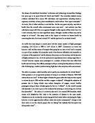
About 90% of those infected with Mycobacterium tuberculosis have , latent TB infection (sometimes called LTBI), with only a 10% lifetime chance that a latent infection will progress to TB disease.However, if untreated, the death rate for these active TB cases is more than 50%. [1][7]
TB infection begins when the mycobacteria reach the , where they invade and replicate within the of alveolar . The primary site of infection in the lungs is called the , and is generally located in either the upper part of the lower lobe, or the lower part of the . Bacteria are picked up by , which do not allow replication, although these cells can transport the bacilli to local () . Further spread is through the bloodstream to other tissues and organs where secondary TB lesions can develop in other parts of the lung (particularly the apex of the upper lobes), peripheral lymph nodes, kidneys, brain, and bone. All parts of the body can be affected by the disease, though it rarely affects the , , and . [4][5]
Tuberculosis is classified as one of the inflammatory conditions. , , and are among the cells that aggregate to form a , with surrounding the infected macrophages. The granuloma functions not only to prevent dissemination of the mycobacteria, but also provides a local environment for communication of cells of the immune system. Within the granuloma, T lymphocytes (CD4+) secrete such as , which activates macrophages to destroy the bacteria with which they are infected. T lymphocytes (CD8+) can also directly kill infected cells.
Importantly, bacteria are not always eliminated within the granuloma, but can become dormant, resulting in a latent infection. Another feature of the granulomas of human tuberculosis is the development of cell death, also called , in the center of . To the naked eye this has the texture of soft white cheese and was termed . [6]
If TB bacteria gain entry to the bloodstream from an area of damaged tissue they spread through the body and set up many foci of infection, all appearing as tiny white tubercles in the tissues. This severe form of TB disease is most common in infants and the elderly and is called . Patients with this disseminated TB have a fatality rate of approximately 20%, even with intensive treatment. [12]
In many patients the infection waxes and wanes. Tissue destruction and necrosis are balanced by healing and . Affected tissue is replaced by scarring and cavities filled with cheese-like white necrotic material. During active disease, some of these cavities are joined to the air passages and this material can be coughed up. It contains living bacteria and can therefore pass on infection. Treatment with appropriate kills bacteria and allows healing to take place. Upon cure, affected areas are eventually replaced by scar tissue [11]
- Pathogenesis of TB infection and disease.
6.3 Diagnosis of pulmonary tuberculosis
Tuberculosis is diagnosed definitively by identifying the causative organism () in a clinical sample for example, sputum or pus. When this is not possible, a probable diagnosis may be made using imaging (X-rays or scans) and/or a .[9]
The main problem with tuberculosis diagnosis is the difficulty in culturing this slow-growing organism in the laboratory which it may take 4 to 12 weeks for blood or sputum culture. A complete medical evaluation for TB must include a medical history, a physical examination, a , microbiological smears and cultures. It may also include a a test. The interpretation of the tuberculin skin test depends upon the person's risk factors for infection and progression to TB disease, such as exposure to other cases of TB or immunosuppression.[10]
Currently, latent infection is diagnosed in a non-immunized person by a tuberculin skin test, which yields a delayed hypersensitivity type response to made from M. tuberculosis.Those immunized for TB or with past-cleared infection will respond with delayed hypersensitivity parallel to those currently in a state of infection, so the test must be used with caution, particularly with regard to persons from countries where TB immunization is common. Tuberculin tests have the disadvantage in that they may produce false negatives, especially when the patient is co-morbid with , Hodgkins lymphoma, malnutrition, or most notably active tuberculosis disease. New TB tests are being developed that offer the hope of cheap, fast and more accurate TB testing. These include detection of bacterial DNA, and assays to detect the release of in response to mycobacterial proteins such as . These are not affected by immunization or , so generate fewer results. The development of a rapid and inexpensive diagnostic test would be particularly valuable in the developing world. [12]
- Treatment of pulmonary tuberculosis
Current recommendations for the treatment of pulmonary tuberculosis include a 6-month course of isoniazid (INH) and rifampin, supplemented during the first 2 months with pyrazinamide. Ethambutol (or streptomycin in children too young to be monitored for visual acuity) may need to be included in the initial regimen until the results of drug susceptibility studies are available. Drug susceptibility studies may not be required if the risk of drug resistance is not significant. Significant risk factors include residence in a community with greater than 4% primary resistance to INH, history of previous treatment with antituberculosis drugs, history of exposure to a drug-resistant case, and origin in a country with a high prevalence of drug resistance. The purpose of this recommendation is to decrease the development of multidrug-resistant (MDR) tuberculosis in areas in which primary INH resistance is increased.[3]
Another treatment option is a 2-month regimen of INH, rifampin, and pyrazinamide daily, followed by 4 months of INH and rifampin twice a week. Effective treatment of hilar adenopathy when the organisms are fully susceptible is a 9-month regimen of INH and rifampin daily or a 1-month regimen of INH and rifampin once a day followed by 8 months of INH and rifampin twice a week.[4]
Because poor adherence to these regimens is a common cause of treatment failure, directly observed therapy (DOT) is recommended for treatment of tuberculosis. DOT means a health care provider or other responsible person must watch the patient ingest the medications. Intermittent regimens should be monitored by DOT for the duration of therapy because poor compliance may result in inadequate drug delivery. [4]
Another initiative recently launched by the WHO is the DOTS-plus strategy, which is based on finding appropriate treatment strategies for MDR tuberculosis and drug susceptibility testing, as well as judicious usage of second-line drugs. It also focuses on community involvement and a good recording and reporting system. [5]
6.5 Chemotherapy Today
Following streptomycin, p-aminosalicylic acid (1949), isoniazid (1952), pyrazinamide (1954), cycloserine (1955), ethambutol (1962) and rifampin (rifampicin; 1963) were introduced as anti-TB agents. Aminoglycosides such as capreomycin, viomycin, kanamycin and amikacin, and the newer quinolones (e.g. ofloxacin and ciprofloxacin) are only used in drug resistance situations. Combinations of a B-lactam antibiotic with a B-lactamase inhibitor enhance treatment effectiveness, but the newer drugs, including the macrolides, have not received much clinical testing.[4][5]
Two properties of anti-TB drugs are important: antibacterial activity, highest in
- isoniazid
- rifampin
- streptomycin
and their capacity to inhibit the development of resistance, the most effective drugs being
- isoniazid
- rifampin
- ethambutol
With the proper four drug regimen, there should be a rapid clinical improvement and a significant fall in the bacterial count. After a month, the patient should be afebrile, feel well and have regained weight. Coughing and sputum should have diminished and improvements will be visible on the X-rays. Although bacteria will still be present in the smears, they will become more and more difficult to culture. Improvements will be visible on the X-rays for three to four months. If the disease was initially severe, though, the end of treatment may not be reached for a year.[14]
The absence of radiological improvement in the first three months should be grounds for concern and indicate that a change in therapy is needed. Patient compliance and the bacteria's drug sensitivity should be reevaluated. Relapses usually occur within six months of the end of treatment, and in most cases are due to poor patient compliance. Patient compliance must be monitored throughout treatment; this is done at the National Tuberculosis Center through .
When TB becomes active again in a previously treated patient, there is a high chance that the bacteria will now be drug resistant. Any current therapy must be suspended until multiple drugs are found to which the pathogen is fully sensitive, and treatment can be resumed with the addition of these drugs to the original regimen. Never add a single drug to a failing regimen. If the microorganism is resistant to the standard drugs, then it will be necessary to administer more toxic medications such as
- ethionamide
- protionamide
- pyrazinamide
- cycloserine
- capreomycin
- viomycin
- kanamycin
Conclusion
The most common area of the body that TB affects is the lungs. This is known as pulmonary tuberculosis.
Pulmonary tuberculosis is a treatable disease if caught early. It does not discriminate, thus anyone, especially individuals with compromised immune systems should adopt healthy lifestyles to protect themselves against TB infection.
TB is not caused by perspiration drying on one’s back, by over-exertion or fatigue TB is not caused by smoking or pollution, although these clearly damage the lungs.
TB is not caused by poor nutrition, although this could make a person who inhales a TB bacterium more likely to develop TB disease. TB is not transmitted through food or drinks or using other utensils. TB is acquired through exposure to someone sick with active TB of the lungs.
Mild symptoms of primary pulmonary tuberculosis and brief, often no obvious signs, many patients were unknowingly spent, and showed that PPD.
TB prevention and control takes two parallel approaches. In the first, people with TB and their contacts are identified and then treated. Identification of infections often involves testing high-risk groups for TB. In the second approach, children are to protect them from TB. No is available that provides reliable protection for adults. However, in tropical areas where the levels of other species of mycobacteria are high, exposure to gives some protection against TB.
Since humans are the only host of Mycobacterium tuberculosis, eradication would be possible. This goal would be helped greatly by an effective vaccine.
Treatment for TB uses to kill the bacteria. Effective TB treatment is difficult, due to the unusual structure and chemical composition of the mycobacterial cell wall, which makes many antibiotics ineffective and hinders the entry of drugs.
Progression from TB infection to TB disease occurs when the TB bacilli overcome the immune system defenses and begin to multiply. In primary TB disease—1–5% of cases—this occurs soon after infection. However, in the majority of cases, a occurs that has no obvious symptoms. These dormant bacilli can produce tuberculosis in 2–23% of these latent cases, often many years after infection. The risk of reactivation increases with immunosuppression, such as that caused by infection with . In patients co-infected with M. tuberculosis and HIV, the risk of reactivation increases to 10% per year.
REFERENCES
Books
-
Frieden TR, Sterling TR, Munsiff SS, Watt CJ, Dye C: Tuberculosis (Seminar). The Lancet 2003, 362(9387):887-899.
- Harries AD, Mphasa NB, Mundy C, Banerjee A, Kwanjana JH, Salaniponi FM. Screening tuberculosis suspects using two sputum smears. Int J Tuberc Lung Dis 2000;4(1):36-40
- Harries AD, Maher D (eds).The diagnosis of tuberculosis in adults: A clinical manual 1996. WHO: 37-38.
-
World Health Organization – Geneva 2003: Treatment of TB: Guidelines for National Programmes.
- Harries AD, Maher D (eds). Standardized TB case definitions and treatment categories: A clinical manual 1996. WHO: 79-82.
-
Kumar, Abbas, Fausto, Mitchelle, Robbins Basic Pathology: Tuberculosis. 8th edition: 516- 523.
Website
-
Starck, Joakim; Kallenius, Gunilla; Marklund, Britt-inger; Andersson,Dan; & Akerlund, Thomas.(2004). Comparative Proteome Analysis of Mycobacterium Tuberculosis grown under Aerobic and Anaerobic Conditions. Microbiology 150: 3821-3829. Doi: 10.1099/mic.0.27284-0
-
Tuberculosis Introduction :
-
Diagnostic Standards and Classification of Tuberculosis, reviewed from
-
Sepkowitz K. Concentric Circles and Knockout Mice: Advances in TB Detection, Diagnosis, and Treatment. 40th Interscience Conference on Antimicrobial Agents & Chemotherapy. search and web pag
- Tuberculosis. WHO Fact Sheet Number 104, Revised April 2000. WHO/OMS web page 2000.
-
Diagnostic Standards and Classification of Tuberculosis, reviewed from
-
Tuberculosis , reviewed from
-
DOTS, reviewed from
-
Summary on TB site: