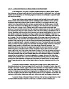
The features of the Gitelman-Bartter syndrome, including hypokalaemic alkalosis, hypermagnesiuria, and hypocalciuria, are mimicked by thiazides. Thiazide diuretics interfere with electrolyte transport in the early distal tubule, so that this nephron segment is involved in patients with Gitelman-Bartter syndrome. These patients only have a partial diuretic response to thiazide diuretics contrasting with a normal response to frusemide. The thiazide-sensitive sodium-clacium cotransporter (TSC) of the early distal tubule has been cloned. With the help of this probe, Simon et al have demonstrated that the hypokalaemic-hypomagnesaemic tubular disorder with hypocalciuria is linked to the gene encoding the TSC located on chromosome 16q. Various mutations in the TSC gene have already been identified.
The pathophysiology of GS has been clearly outlined by Bhandari. The defect in the TSC leads to NaCl wasting and hypovolemia which stimulates the rennin-angiotensin-aldosterone system, and causes an increase in apical sodium reabsorption and stimulation of the basolateral Na+/k+-ATPase. The increased aldosterone levels also stimulate cortical and medullary collecting ducts H+/ATPase pumps leading to an increased apical hydrogen ion secretion. K+ and H+ ion excretion increases as K+ enters from the basolateral membrane via the Na+/K+-ATPase pumps, resulting in hypokalemic metabolic alkalosis. The resultant low intracellular Na+ increases distal convoluted tubule calcium reabsorption via basolateral Na+/Ca++ exchangers, causing hypocalciuria. Mg++ loss, via apical Mg++/Na++ exchangers, increases due to the net negative transepithelial potential. This reduced Mg++ may also stimulate parathormone release, which further increases Ca++ reabsorption. Hypermagnesuria may be caused balso by associated hypokalemia, metabolic alkalosis, or low renal Mg threshold. Hypomagnesia and hypocalciuria may also occur following the administration of thiazide diuretics which inhibit the distal luminal Na-Cl cotransporter.
Renal tubular defects in the Na-K-2Cl transporter prevents urine calcium reabsorption in the thick ascending limb of Henle. Continued reabsorption and secretion of the potassium ions into the lumen of the TAL promotes reabsorption of the positive calcium ions through paracellular channels. Excessive urine calcium excretion may be one factor in the nephrocalcinosis observed in these patients.
Calcium is usually reabsorbed in the distal convoluted tubule. Theoretically, chloride is reabsorbed through the thiazide-sensitive Na-Cl cotransporter and transported out of the cell through a basolateral chloride channel, reducing intracellular chloride concentration. The net effect is increased activity of the voltage-dependent calcium channels and enhanced electrical gradient for calcium reabsorption from the lumen.
Bartter syndrome is caused by Na+ and Cl- wasting in the thick ascending limb of the loop of Henle. Abnormalities in NKCC2, ROMK, and ClC-Kb have all been reported. Frameshift, nonsense, or missense mutations of the gene for NKCC2 have been found in affected patients of five families. These mutant alleles cause loss in NKCC2 function leading to salt wasting in the loop of Henle. Reduced Cl- absorption in the loop of Henle inhibits the voltage-driven paracellular reabsorption of calcium and magnesium, causing hypercalciuria and hypermagnesuria. Prostaglandins also contribute to urinary calcium and magnesium losses by increasing bone reabsorption. Hypercalciuria is an important feature of Bartter syndrome and is associated with development of nephrocalcinosis. Enhanced magnesium reabsorption in the distal nephron may compensate for proximal hypermagnesuria, reduced magnesium wasting and preventing significant hypomagnesemia. Increased prostaglandin activity in Bartter syndrome may stimulate the hydroxylation of vitamin D, which would result in an increase in serum 1,25 (OH)2D concentration. This, in turn, can increase calcium reabsorption, promoting hypercalciuria and nephrocalcinosis.
In Gitelman syndrome, dysfunction of the Na-Cl cotransporter leads to hypocalciuria and hypomagnesemia. How this occurs is not clear. Simon et al showed complete linkage of Gitelman’s syndrome to the locus encoding the thiazide-sensitive Na-Cl cotransporter. The reasons for the high levels of urinary magnesium and low levels of urinary calcium are not clearly understood. It is suggested that continued efflux of intracellular Cl- through basolateral CIC-Kb might promote calcium reabsorption in the cells of the distal convoluted tubule, thus accounting for hypocalciuria. This segment of the nephron also reabsorbs about 5% of filtered magnesium. Loss of function of the NCCT might inhibit magnesium absorption by a sodium-dependent mechanism.
IN complex tubular disorders the more downstream part of the tubule may modulate or compensate for the upstream tubular dysfunction.
Hypomagnesemia can cause hypocalcemia because it interferes with the calcium-elevating effects of parathyroid hormone (PTH).
The kidney occupies a central role in magnesium balance. Factors that modulate and affect renal magnesium excretion can have profound effects on magnesium balance. In turn, magnesium balance affects numerous intracellular and systemic processes.
A major phenotypic difference between Bartter’s and Gitelman’s syndromes involves urinary calcium excretion. The hypercalciuria of Bartter’s syndrome is believed to result largely from dysfunction of thick ascending limb cells. Calcium absorption along the loop of Henle is largely passive, paracellular, and driven by the lumen-positive transepithelial voltage that is generated by Na-K-2Cl cotransport and luminal K recycling. When Na-K-2Cl cotransport is reduced or blocked by loop diuretics or genetic abnormality, the lumen-positive voltage declines or approaches zero, and calcium reabsorption declines. Another cause may also contribute to calcium wasting. Rates of sodium and calcium transport by distal tubules tend to correlate inversely. NaCl transport by DCT cells will be increased in patient’s with Bartter’s syndrome. This raises the intracellular Cl activity of DCT cells, causing depolarisation and inhibition of the apical calcium channel. This would be predicted to impair distal calcium reabsorption, providing a distal contribution to calcium wasting and nephrolithiasis.
Patients with Gitelman’s syndrome, in contrast, show hypocalciuria, which resembles the clinically useful effect of DCT diuretics to reduce urinary calcium excretion. The mechanisms of hypocalciuria are well established. First, mild contraction of the extracellular fluid volume will increase calcium reabsorption along the proximal tubule. The reduction in NaCl entry into DCT cells stimulates transepithelial calcium transport. The intracellular Cl concentration declines, causing a lower intracellular Cl activity, which hyperpolarizes the cell. This hyperpolarisation activates a distinctive calcium channel that is expressed by DCT cells, thus increasing apical calcium entry.
For the increase in apical calcium entry to result in increases in transepithelial calcium transport, calcium movement from lumen to cell must be balanced by calcium movement from cell to interstitium and blood. The increase in cellular calcium consequent to increased luminal calcium entry will stimulate calcium efflux via the basolateral Na/Ca exchanger and the Ca-ATPase, but other factors contribute as well. First, inhibition or absence of apical NaCl entry reduces the intracellular Na activity, which stimulates 3Na/Ca exchange. Second, the hyperpolarisation stimulates Na/Ca exchange because this transport protein operates in an electrogenic mode, carrying 3Na ions into the cell for each calcium ion extruded.
Figure 1: Electrolyte transport in the distal convoluted tubule.
Figure 2: Pathways and mediators involved in Na+ reabsorption.
Figure 3: Features differentiating Bartter and Gitelman syndromes.
Figure 4: Mechanism of electrogenic chloride reabsorption in the medullary thick ascending limb of the loop of Henle. Passive paracellular absorption of calcium and magnesium depends on the transepithelial potential difference generated by electrogenic chloride transport.Hypercalciuria and hypermagnesiuria may be caused by impaired chloride reabsorption.
Figure 5: The glomerular filtration of magnesium.
Figure 6: The renal handling of magnesium. Magnesium is filtered at the glomerulus, with the ultrafilterable fraction of plasma magnesium entering the proximal convoluted tubule (PCT). At the end of the PCT, the magnesium concentration is approximately 1.7 times the initial concentration of magnesium and about 20% of the filtered magnesium has been reabsorbed. Magnesium reabsorption occurs passively through paracellular pathways. Hydrated magnesium has a very large radius that decreases its intercellular permeability in the PCT when compared with sodium. The smaller hydrated radius of sodium is 50% to 60% reabsorbed in the PCT. No clear evidence exists of transcellular reabsorption or secretion of magnesium within the mammalian PCT. In the pars recta of the proximal straight tubule (PST), magnesium reabsorption can continue to occur by way of passive forces in the concentrating kidney. In states of normal hydration, however, very little magnesium reabsorption occurs in the PST. Within the thin descending limb of the loop of Henle, juxtamedullary nephrons are capable of a small amount of magnesium reabsorption in a state of antidiuresis or magnesium depletion. This reabsorption does not occur in superficial cortical nephrons. No data exist regarding magnesium reabsorption in the thin ascending limb of the loop of Henle. No magnesium reabsorption occurs in the medullary portion of the thick ascending limb of the loop of Henle; whereas nearly 65% of the filtered load is absorbed in the cortical thick ascending limb of the loop of Henle in both juxtamedullary and superficial cortical nephrons. A small amount of magnesium is absorbed in the distal convoluted tubule. Magnesium transport in the connecting tubule has not been well quantified. Little reabsorption occurs and no evidence exists of magnesium secretion within the collecting duct. Normally, 95% of the filtered magnesium is reabsorbed by the nephron. In states of magnesium depletion the fractional excretion of magnesium can decrease to less than 1%; whereas magnesium excretion can increase in states of above-normal magnesium intake, provided no evidence of renal failure exists.
Figure 7: Magnesium reabsorption in the cotical thick ascending limb (cTAL) of the loop of Henle. Most magnesium reabsorption within the nephron occurs in the cTAL owing primarily to voltage-dependent Mg flux through the intercellular tight junction. Transcellular Mg movement occurs only in response to cellular metabolic needs. The sequence of events necessary to generate the lumen-positive electrochemical gradient that drives Mg reabsorption is as follows: 1) A basolateral sodium-potassium-adenosine triphosphate (Na+-K+-ATPase) decreases intracellular sodium, generating an inside-negative electrical potential difference; 2) Intracellular K is extruded by an electroneutral K-Cl cotransporter; 3) Cl is extruded by way of conductive pathways in the basolateral membrane; 4) The apical-luminal Na-2Cl-K (furosemide-sensitive) cotransport mechanism is driven by the inside-negative potential difference and decrease in intracellular Na; 5) Potassium is recycled back into the lumen by way of an apical K conducitve channel; 6) Passage of approximately 2 Na molecules for every Cl molecule is allowed by the paracellular pathway (intercellular tight junction), which is cation permselective; 7) Magnesium reabsorption occurs passively, by way of intercellular channels, as it moves down its electrical gradient.
Figure 8: Renal magnesium wasting. Mg is normally reabsorbed in the proximal tubule (PT), cTAL and DCT. Volume expansion and osmotic diuretics inhibit PT absorption of magnesium. Several renal diseases and electrolyte disturbances inhibit Mg reabsorption in both the PT and cTAL owing to damage to the epithelial cells and the intercellular tight junctions, plus disruption of the electrochemical forces that normally favour magnesium reabsorption. Many drugs and toxins directly damage the cTAL. Thiazides have little direct effect on magnesium reabsorption; however, the secondary hyperaldosteronism and hypercalcemia effect Mg reabsorption in CD and/or cTAL. Aminoglycosides accumulate in the PT, which affects sodium reabsorption, also leading to an increase in aldosterone. Aldosterone leads to volume expansion, decreasing magnesium reabsorption. Parathyroid hormone has the direct effect of increasing magnesium reabsorption in cTAL; however, hypercalcemia offsets this tendency. Thyroid hormone increases Mg loss. Diabetes mellitus increases Mg loss by way of both hyperglycaemic osmotic diuresis and insulin abnormalities (deficiency and resistance), which decrease Mg reabsorption in the PCT and cTAL, respectively.