This is a hard question to answer because good studies are just now being conducted (3). Currently, there is limited evidence to show that human skeletal muscle switches fiber types from "fast" to "slow" due to training (4). Researchers have demonstrated a fast-to-slow fiber transformation in animal skeletal muscle, and the human studies are showing similar outcomes. There is decent evidence that pure fast (Type IIb) fibers can transition to "hybrid" (Type IIa) fibers with chronic endurance training.
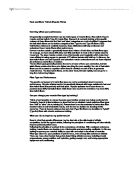
What can I do to improve my performance?
Keep in mind that genetic differences may be dramatic at the elite levels of athletic competition, but for the typical athlete, following the principles of conditioning will dramatically improve personal performance.
Following the principle of overload is the cornerstone of training. With consistent endurance training muscle fibers can develop more mitochondria and surrounding capillaries. In this way training improves your muscle's ability to cope with and adapt to the stress of exercise.
Fiber type alone is a poor predictor of performance, even among elite endurance athletes. There are many other factors that go into determining athletic success, including mental preparedness, proper nutrition and hydration, getting enough rest, and having appropriate equipment and conditioning.
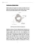
The background image shows the internal structure of a muscle fibre (cell).
- Myofibrils are the protein rods which are made to slide past each other when a muscle actively contracts
- Sarcoplasmic reticulum stores calcium ions and releases it to initiate contraction and pumps it in to end contraction.
- Terminal cisternea are specialised regions of the sarcoplasmic reticulum which make contact with the transverse tubules. Calcium ions is released from this region onto the contractile filaments. Calcium ions trigger active sliding of the filaments, which produces muscle shortening.
- Opening of the transverse tubules to the space outside of the muscle cell. Electrical signals travel from the outside surface of the muscle deep into the muscle fibre down the transverse tubules.
- Myoplasm is the surface membrane of the muscle cell. This membrane carries electrical signals (the action potential) along the muscle fibre. The action potential travels into the muscle fibre down tranverse tubules.
Muscle Fibre Types
Skeletal muscle fibres are not all the same.
Traditionally, they were categorised depending on their varying colour.
Red Fibres:
Those containing high levels of myoglobin and oxygen storing proteins had a red appearance. Red muscle fibres tend to have more mitochondria and blood vessels than the white ones.
White Fibres:
Those with a low content had a white appearance.
To further confuse the issue skeletal muscle fibres are also classified, depending on their twitch capabilities, into fast and slow twitch.
Fast Twitch:
Some authors define a fast twitch fibre as one in which the myosin can split ATP very quickly.
However, fast twitch fibres also demonstrate a higher capability for electrochemical transmission of action potentials and a rapid level of calcium release and uptake by the sarcoplasmic reticulum. The fast twitch fibres rely on a well developed, short term, glycolytic system for energy transfer and can contract and develop tension at 2-3 times the rate of slow twitch fibres.
Slow Twitch:
The slow twitch fibres generate energy for ATP re-synthesis by means of a long term system of aerobic energy transfer. They tend to have a low activity level of ATPase, a slower speed of contraction with a less well developed glycolytic capacity. They contain large and numerous mitochondria and with the high levels of myoglobin that gives them a red pigmentation they have been demonstrated to have high concentration of mitochondrial enzymes, thus they are fatigue resistant.
The 2 main categories of muscle fibres become 3 when we split the white muscle fibres into 2 sections.
So we expand further:
Type I
Red fibres.
Slow oxidative (also called slow twitch or fatigue resistant fibres).
Contain:
Large amounts of myoglobin.
Many mitochondria.
Many blood capillaries.
Generate ATP by the aerobic system, hence the term oxidative fibers.
Split ATP at a slow rate.
Slow contraction velocity.
Resistant to fatigue.
Found in large numbers in postural muscles.
Needed for aerobic activities like long distance running.
Type IIa
Red fibres.
Fast oxidative (also called fast twitch A or fatigue resistant fibers).
Contain:
Large amounts of myoglobin.
Many mitochondria.
Many blood capillaries.
High capacity for generating ATP by oxidation. Split ATP at a very rapid rate and, hence, high contraction velocity.
Resistant to fatigue but not as much as slow oxidative fibres.
Needed for sports such as middle distance running and swimming.
Type IIb
White.
Fast glycolytic (also called fast twitch B or fatigable fibres).
Contain:
Low myoglobin content.
Few mitochondria.
Few blood capillaries.
Large amount of glycogen.
Split ATP very quickly.
Fatigue easily.
Needed for sports like sprinting.
Individual muscles are a mixture of 3 types of muscle fibres (type 1 and type 2a and b), but their proportions vary depending on the action of that muscle.
It must be remembered that skeletal muscles, although a mixture, can only have one type of muscle fibre within a motor unit. This is demonstrated if we look at contractions.
E.g. If a weak contraction is needed only the type 1 motor units will be activated. These fibres are used mainly for endurance activities.
If a stronger contraction is required the type 2a fibres will be activated or used to assist the type 1 fibres.
Maximal contractions facilitate the use of type 2b fibres which are always activated last. These fibres are used during ballistic activities but tire easily.
With advanced EMG techniques it is possible to look at which muscle fibres are recruited when performing an exercise/test.
The total number of skeletal muscle fibres has traditionally been thought not to change.
It is believed there are no sex or age differences in fibre distribution, however, relative fibre types vary considerably from muscle to muscle and person to person.
Sedentary men and women (as well as young children) have 45% type 2 and 55% type 1 fibres.
People at the higher end of any sport tend to demonstrate patterns of fibres distribution e.g. endurance athletes show a higher level of type 1 fibres.
Sprint athletes, on the other hand, require large numbers of type 2 b fibres.
Middle distance event athletes show approximately equal distribution of the 2 types. This is also often the case for power athletes such as throwers and jumpers.
It has been suggested that various types of exercise can induce changes in the fibres of a skeletal muscle.
It is thought that if you perform endurance type events for a sustained period of time, some of the type 2b fibres transform into type 2a fibres. However, this is argued about.
It may well be that the type 2b fibres show enhancements of the oxidative capacity after high intensity endurance training which brings them to a level at which they are able to perform oxidative metabolism as effectively as slow twitch fibres of untrained subjects. This would be brought about by an increase in mitochondrial size and number and the associated related changes not a change in fibre type.
Muscle Contraction: Sliding Filaments
(A) The myosin and actin filaments of a sarcomere overlap with the same relative polarity on either side of the midline. Recall that actin filaments are anchored by their plus ends to the Z disc and that myosin filaments are bipolar. (B) During contraction, the actin and myosin filaments slide past each other without shortening. The sliding motion is driven by the myosin heads walking toward the plus end of the adjacent actin filament.