How does the skeletal system help Sports performance?
How does the skeletal system help . Sports performance?The skeletal system plays a very important part to helping sporting performance. It acts as a framework for muscle attachment and some bones protect vital organs, e.g. the cranium protects the brain. Other bones produce red and white blood cells. The Skeletal system also supports the body and determines shape. There are 4 different types of bones. These are; Long bones, Short bones, flat bones and irregular bones. Long bones are longer in length than width and are cylindrical in shape; these are associated with movement, e.g. Humerus and Tibia. Short bones are there for strength but have limited movement. They tend to be squarish in shape, E.g. Tarsal and carpals. Flat bones are the bones that enclose our vital organs.
An example of a flat bone is ribs or scapula. Irregular bones are bones that have no specific shape, but are used for muscle attachment in the body and also, importantly, give us our shape. An example of an irregular bone is the Patella or vertebrae. The vertebral column also is a very vital part to our body as it supports our body, allowing us to stand up, bend flex and twist. There are 5 sections to the vertebral column. The cervical, which consists of 7 vertebrae. These are the smallest vertebrae and are found in the neck. Then there ...
This is a preview of the whole essay
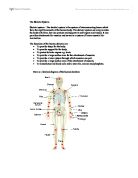
An example of a flat bone is ribs or scapula. Irregular bones are bones that have no specific shape, but are used for muscle attachment in the body and also, importantly, give us our shape. An example of an irregular bone is the Patella or vertebrae. The vertebral column also is a very vital part to our body as it supports our body, allowing us to stand up, bend flex and twist. There are 5 sections to the vertebral column. The cervical, which consists of 7 vertebrae. These are the smallest vertebrae and are found in the neck. Then there is the Thoracic. This consists of 12 vertebrae. The 12 pairs of ribs join on to the thoracic vertebrae. They are much larger than the cervical vertebrae. The lumbar region, which follows after the Thoracic has 5 vertebrae. This is the largest and strongest part of the vertebral column. They are found in between the gap of the ribs and pelvic girdle so therefore is probably the region vulnerable to the most injury. The sacrum consists of 5 fused vertebrae, which form the rear part of the pelvic girdle. Finally, the Coccyx. Consisting of 4 fused vertebrae. It is thought to be the remains of a tail, which may once have existed. The diagram on the left shows the bones in the body, which will give you a visual example of the long, short, flat and irregular bones. Also, there are several different joints that join two or more bones together. However, there are joints called Synovial joints, which differ, from normal joints. A Synovial joint is lubricated with a clear blood plasma from the capillaries and filtered through a membrane around it. The fluid works with pads of fat which is stored around the joint to act as a cushion between bones. The fluid also helps maintain the cartilage, in and around the joint. Below are the parts of a Synovial joint. It is mainly the Epiphysis of the long bones which form the Synovial joints. As well as joints, there are ligaments. They connect bone to bone. They are flexible with very strong bundles of parallel fibres. They stop the bones dislocating by restricting movement. Throughout the years, bones grow and change. Especially through out childhood. This is called Ossification. At Birth, all the bones are just cartilage, or a cartilage model. Then, the Periosteal collar appears, which is the start of the cartilage in the shaft of the bone calcifies. As the cartilage dies, gaps are filled with blood vessels, called Osteoblasts and Osteoclasts. Ossification occurs in three parts of the bone. This is the 2 Epiphysis and the Diaphysis. By mid 20’s, all bones will be at there final strength and length. Throughout this piece, I will be focusing on the sport “netball” and explaining the bones and joints used within this sport. There are several different passes in netball, but I am going to focus more into the depth of the chest pass. The chest pass is the most accurate and strongest way of getting the ball around the court. The ball is held with your hands shaped in a “w”, which is done by bringing your thumbs together and spreading your fingers out. Keep your arms close to your body and have the ball against your chest. It should look something like this: Or Within this position, one leg, preferably your strongest should be slightly behind for better balance. Then, push the ball away from you, stepping forward with the foot that was once behind, to create more power when pushing the ball through. Keeping your elbows close to your body, push through with the ball. As you release the ball, straighten your arms and fingers. Keep your wrists pointed upwards to help fully extend your arms. It should look something like this: Through this pass, Leg and arm bones and joints will have been used. The bones used in the arms will be the Humerus, Ulna, Radius, Carpals, Metacarpals and Phalanges. The joints used in the arms are called pivot (between radius and ulna) and hinge (between the radius ulna and humerus). The bones used within the leg and foot are femur, tibia, fibula tarsal and metatarsals. Long bones play the most important part out of all of the bones, as they allow the most movement. Bones do play a very important part when playing sport, and has many functions, which are protecting you and preventing injury. Netball is a good example of the use of several bones and joints around the body. By Sabrina Murphy.