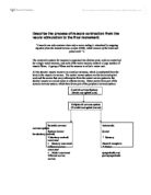
Uncontrolled variable: Length and width of Chicken muscle fiber
Materials:
- Muscle fibers
- Millimeter graph paper
- 3 glass slides
- 3 droppers
- Distilled water
- Glucose solution
- ATP solution
- Stop clock
- Blunt mounted needles
- Petri dish
Method:
1. Place the muscle fiber on a glass slide. Place the slide on the piece of graph paper.
2. Use the mounted needles to straighten the fibre (the lines on the graph paper should help)
3. Measure and record the length of the fibre.
4. Put 3 drops of water on the fibre so that the whole length of the fibre is covered. Leave for 2 minutes. Drain the water off into the Petri dish. Empty the dish.
5. Repeat steps 2 (if necessary) and 3.
6. Repeat step 4 using glucose solution instead of water.
7. Repeat steps 2 (if necessary) and 3.
8. Wash and drain away the glucose from the muscle with several drops of water.
9. Repeat steps 4 using ATP solution instead of water.
10. Repeat steps 2 (if necessary) and 3.
Data Collection:
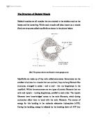
The figures above are not 100% accurate. This is because eyes of human beings are not 100% accurate. Therefore a scope of 0.5mm has to be taken into account when looking at the figures.
Conclusion and Evaluation:
The investigation was carried out successfully. We had varied results in the class, but they were not very far off from each other in most cases. My hypothesis was correct for the chicken muscle fiber. Looking at the average figures for the test carried out it is seen that when water was added there was a slight expansion in one case of the chicken, when glucose was added, there was a small contraction as suspected in the chicken muscle. When ATP was added the muscle fibers contracted significantly. These results can be explained by the fact that when water is added to muscles, due cells give way to outside water pressure and let in the water, the expanding the muscle. Glucose, a substance the body needs to provide energy; I though would contract the muscles because I thought the cells would be much more active when presented with this solution. However this was seen in the chicken muscle. When ATP was added there was significant contraction. This is because ATP is the final form on energy that the body uses. It is therefore that the muscles contract because they are much more active when there is a higher concentration of ATP.
Certain inaccuracies throughout this experiment have to be taken into consideration when looking at the results. Firstly the pieces of muscle fibres tested were not of the same length, width and this could have caused differences in the reactions to the solutions presented. Thirdly when using litmus paper to absorb the added solution, this procedure sometimes this was done very carefully, leaving more solution on some tests and less on others. This could cause certain changes in the results as some tests had more solution on the than others. Finally, because the eye is not 100% correct, and because the graph paper is not 100% accurate, when looking at the fibres to check for changes; mistakes could have been made in noting these changes.
The litmus paper could be used in a more efficient way, if I placed it on the muscle fibre for the same amount of time on each test. In addition to this I could have used more accurate graph paper to give more accurate results. Also the chicken that we used was probably killed since a few days. Many cells could have died in these few days, and so become less reactive to the solutions that were added. If the chicken was fresher, say just cut the same day; I think I would have more accurate results because the cells would react faster to the solutions. The pieces cut out of the chicken could have more accurate by using a computer controlled cutting machine.