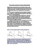
Cellulose forms crystalline structures, using hydrogen bonding both inside the molecule and between the molecules. Water is a catalyst for the formation of these crystals, as the water molecules help the cellulose molecules line up. This make it almost completely insoluble in water under normal conditions, unlike amylose molecules, or for that matter, glycogen.
The structure of glycogen has been known and agreed upon by all the academic literature, since the 1970s. Glycogen is a water soluble molecule that is formed around a protein core, of a molecule called glycogenin. Radiating out from the centre are polysaccharide chains that bifurcate. The total molecule is therefore spherical in appearance, a sort of ‘hairy sphere’. The highly branched structure is important for the metabolism of the glycogen molecule, as it maximises the surface area available for reaction, while its spherical shape allows the molecule to travel unimpeded through the blood, and not ‘snag’.
Aim: To compare the hydrolysis of glycogen, starch and cellulose. A spectrophotometer will be used and this is a form of colour measurement and it assigns numerical values dependant on the level of absorption of solution in cuvette.
Method: Take 12 test tubes and place in a test tube rack. Pipette 1ml of glycogen into 6 tubes, and 1 ml of other polysaccharide into the remaining 6 tubes. Take a clean pipette and add 1 ml distilled water, then using a 5ml pipette add 2 ml of 2M HCl to each tube.
Cover with parafilm and pierce this with a pin. Place the 12 test tubes in a boiling water bath, making a note of the time. Using the test tube tongs, remove one tube of each polysaccharide solution from the water bath after 10 minutes. Upon removal-using a Pasteur pipette, take one drop of each solution and place in separate wells of the white spotting tile. Add one drop of iodine to each and mix, making note of the colour produced. Neutralise the rest of the solution in the tubes by adding 1ml of 4M NaOH to each. Record the results at every ten minutes interval
For spectrophotometer
Pierce and cover with parafilm each test tube Add 1 ml of DNS put to water bath for 5 minutes. Then allow it to cool room temperature, Prepare a blank by placing 1ml DNS reagent and 2ml distilled water into a cuvette, this will be used to zero the spectrophotometer. When the test tubes are cool pour each solution into a separate cuvette and read the extinction at 540nm against the blank. Repeat this process for each tube at appropriate time intervals.
Results
Graph:
Graph 1a shows how the absorption of cellulose changes over time.
Discussion
The graph shows that glycogen gets hydrolysed faster than cellulose. Therefore we can come to the conclusion that Cellulose is more unwilling to hydrolysis than glycogen. The reason for this can be seen in the structure of cellulose in comparison to glycogen. Some of the reasons are below
- The orientation of the bonds linking the glucose residues, the rings of glucose are arranged in a flip-flop manner. This produces a long, rigid molecule.
- There are no side chains in cellulose as there are in glycogen. The absence of side chains allows these linear molecules to lie close together.
-
Because of the many -OH groups, as well as the oxygen atom in the ring, there are many opportunities for to form between adjacent chains.
The result is a series of stiff, elongated fibrils - the perfect material for building the cell walls of plants. Therefore cellulose is much more resistant to hydrolysis than glycogen. Animals store excess glucose by polymerizing it to form glycogen. The structure of glycogen is similar to that of amylopectin, although the branches in glycogen tend to be shorter and more frequent.
Glycogen is broken back down into glucose when energy is needed (a process called glycogenolysis), Therefore the breakdown on glycogen has to be easy.
Unfortunately I didn’t get the opportunity to use starch in the experiment as there wasn’t enough time available. If I was to repeat the experiment I would have done more tests and a wider range of time, this is to improve the accuracy of my results. I would also use other polysaccharides to see if there is a trend.