Effect of temperature on the enzyme pectinase in fruit juice production.
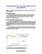
Effect of temperature on the enzyme pectinase in fruit juice production AIM: To investigate the effect of temperature on the work of the enzyme pectinase in fruit juice production. INTRODUCTION It is stated that temperature has an effect on Enzymes. I am going to investigate this fact using my chosen enzyme Pectinase. This enzyme is used in the fruit juice industry and I am going to use this enzyme to explore the question: How Does Temperature Affect Enzymes? BACKGROUND INFORMATION Enzymes are biological catalysts that speed up the rate of a reaction without being changed or used up in any way. They are protein molecules that are tailored to recognize and bind specific reactants and speed their conversion into products. These proteins are responsible for increasing the rates of all of the many thousand of reaction taking place inside cells. Activation energy is the energy barrier over which the molecules in a system must be raised for a reaction to take place. The use of enzymes lowers the activation energy so the reaction can proceed at a faster rate. ( GRAPH TAKEN FROM REFERENCE 7) A number of factors affect the activity of enzymes in speeding conversion of reactants to products. These factors are; . PH: Each enzyme has an optimal pH range that help maintain its normal configuration in an environment which it operates. The tertiary structure of a protein

The Effect of pH on Pectinase
The Effect of pH on Pectinase Hypothesis I plan to investigate the effects of varying pH on the enzyme 'Pectinase'. I will test the yield of apple juice from a set amount of pureed apple at different pH. I think the optimum pH for pectinase is likely to be around pH 5, an acidic pH, as many commercial pectinases are produced from fungi. Before I look at the structure of pectinase, first it would seem sensible to look at the substrate that pectinase will be working on, Pectin. Starch and Pectin are very similar molecules. Starch and Pectin are both polysaccharides. However in Pectin the repeating unit is not glucose as in starch, but galacturonic acid. Galacturonic acid is very similar to glucose, except that the carbon with CH2-OH attached in glucose is replaced by a -COOH in galacturonic acid (shown below). Bonds between carbon 1 of the first galacturonic acid and carbon 4 of the next hold pectin chains together. This sequence continues down the chain. Enzymes are biological catalysts; they alter the speed of reactions without being reactants themselves. There are two theories on how enzymes work. The first is the 'lock and key theory'. This theory suggests that substrate molecule fits perfectly into its specific enzymes active site, and only that enzyme. This can be seen below: The second theory is the 'induced fit theory'. This suggests that the active site on the
Daphnia experiment - Does caffeine affect heart rate?
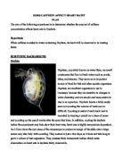
DOES CAFFEINE AFFECT HEART RATE? PLAN The aim of the following experiment is to determine whether the amount of caffeine concentration affects heart rate in Daphnia. Hypothesis When caffeine is added to water containing Daphnia, its heart will be observed to be beating faster. SCIENTIFIC BACKGROUND Daphnia Daphnia, popularly known as water fleas, are small crustaceans that live in fresh water such as ponds, lakes, and streams. They serve as an important source of food for fish and other aquatic organisms. Daphnia are excellent organisms to use in bioassays because they are sensitive to changes in water chemistry and are simple and inexpensive to rise in an aquarium. Daphnia hearts a fairly easily seen but counting the number of beats can be difficult. Counting is easier if each heart beat is recorded by tapping a pencil on a piece of paper and counting up the pencil marks after the specified time. In addition, cooling the daphnia before the experiment may help slow their heart rate: heart rate is highly temperature dependant. An I Cam above the eye-piece of the microscope to project an image of the slide onto a large screen may also help with counting. They mature in just a few days, so it does not take long to grow a culture of test organisms. They possess fairly transparent bodies which make observation on heart rate in daphnia fairly observable.

Relating the structure and function of cell organelles
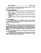
Relate the structure and function of cell organelles Cells are like cities with intricate organelles 'living and working' in it. There are basically two kinds of cells, namely Prokaryotic and Eukaryotic. Both plant cells and animal cells are eukaryotic while prokaryotic cells are simplier organisms that possess non-membrane bounded organelles. In most cells they contains the following organelles which carry out unique functions and allow cells to work properly. To start off with, nucleus is a large roundish organelle enclosed by a double membrane with numerous openings, namely nuclear pores, for nuclear traffic. It contains chromosomes and one or more nucleoli. Nucleolus is a spherical site where ribosomes are formed. Chromosomes contain DNA which tends to be packed in form of chromatin. Only during interphrase (a stage before a cell divides in a cell division process), chromosomes will be unravelled for easier replication. Tiny, hollow cylinders of protein called centrioles form a network of spindle fibres in the nucleus during nuclear division to pull chromosomes apart. The inner membrane of nucleus will break down and allow chromosomes lying freely in cytoplasm. DNA contains the genetic information and control the synthesis of protein. Each cell contains millions of ribosomes. They are very tiny, non-membrane bounded organelles made of protein and RNA which consist of

structural differences between fibrous and globular proteins.
Question: Explain with examples, the structural differences between fibrous and globular proteins. A globular protein has a fixed specific sequence of amino acids that are non-repetitive while a fibrous protein has a repetitive regular sequence of amino acid. For example, haemoglobin, a globular protein is made up of 4 polypeptide chains to form a tetramer (?2?2), composed of two identical alpha-beta (??) dimers. Collagen, a fibrous protein, has a primary structure characterized by a repeating tripeptide sequence of Glycine - X - Y. (X is proline, Y is either hydroxyproline or hydroxylysine) A globular protein has a more compact structure owing to highly contorted pattern of folding, bending and twisting along polypeptide chain to give the protein a spherical 3D shape while a fibrous protein is usually formed with elongated polypeptide chains wrapped around to form multi-molecular paralleled filaments to strands. For example, haemoglobin is a tetramer made up of 4 polypeptide chains of 2? chains and 2? chains. These four subunits are packed to form an overall spherically shaped molecule. However, collagen, a fibrous protein, is formed with three polypeptide chains lie parallel and wind round one another, forming a tropocollagen. The tropocollagen molecules lie side by side and are linked to each other giving a collagen fibril. A globular protein has its length of

Can heart disease be prevented?
Can heart disease be prevented? Preventing heart disease. Something our doctors tell us about all the time, something we all want to do, but what exactly is a "heart disease". How can we prevent it if we don't even know what it is? Every one has heard the terms "heart attack" and "stroke" but hardly anyone knows what they mean. Let's start right at the beginning. A heart disease, medically known as cardiovascular disease, is a disease of the heart and the blood vessels. Most people think only the middle aged and elderly get such diseases but no, cardiovascular diseases can be found in children as young as the age of seven years old. This is strongly liked with the children's lack of exercise and a poor diet. There are many types of cardiovascular diseases of which the major ones are atherosclerosis, coronary, rheumatic, congenital, myocarditis, angina and arrhythmia. Heart disease can arise from congenital defects, infection, narrowing of the coronary arteries, high blood pressure, or disturbances. (1) Atherosclerosis is the thickening of the inner layer of the arterial walls due to the deposit of cholesterol, fibrous tissue, dead muscle cells and blood platelets. This deposit is also known as atheromatous plague or an atheroma. Rheumatic heart disease used to be one of the most serious heart diseases in both children and adolescence as it involves damage to the entire

Beetroot pigments
Assessed Practical Plan I plan to investigate the effects of varying temperature on the movement of pigment molecules across a membrane, travelling out of beetroot cells. Aim: The aim of this investigation is to determine the effect that temperature has on the rate of diffusion of beetroot pigments when slices of beetroot are placed in various temperatures of water. Background Knowledge: The pigments found in beetroot are known as betalain pigments. Some areas of a beetroot may contain more pigment molecules than others. These molecules appear to be polar molecules as they are soluble in water. Molecules which have groups with dipoles are said to be polar. They are usually attached to water molecules. They also have dipoles, and these molecules are hydrophilic, and tend to be soluble in water. The process by which the betalain molecules leave the cell, can be explored by considering the following: * Simple Diffusion * Facilitated Diffusion * Osmosis * Bulk Transport * Active Uptake We can eliminate the possibility that betalain pigments travel by osmosis, since osmosis only involves the movement of water molecules, whereas in this experiment we are researching the movement of beetroot pigment molecules. Also, the possibility of the betalain pigments travelling through the membrane by active uptake or bulk transport can be discarded, as both of these processes

Investigate the water potential of celeriac.
Aim Investigate the water potential of celeriac. Apparatus Celeriac - this is the specimen that we will be finding the water potential of. .00 mol dm-3 sucrose solution - this will be placed in the test tube in which the experiment will take place. We will dilute the solution to produce a range of concentrations. 0.0ml Graduated Pipettes (x2) - I had a choice of 5.0ml, 10.0ml and 20.0ml graduated pipettes. I chose this size pipette because it is time efficient and accurate at the same time. It is an accurate piece of equipment because as opposed to standard pipettes, this graduated pipette has 0.1ml graduations to ensure the utmost accuracy. One pipette will be used for water and one will be used for sucrose solution. 250ml Beakers (x2) - these will be used to hold water and the 1.00 mol dm-3 sucrose solution in a safe environment. Cork Borer - this will be used to cut the precise shape of the celeriac. This will also ensure that the surface area to volume ratio is constant throughout the experiment. Razor - this will be used to cut the pieces of celeriac to a precise length. Ruler - this will be used to measure the length of celeriac that will be used in the experiment. Scales - they will be used to measure the mass of celeriac before and after the experiment with accuracy. Therefore, the mass increase/decrease can be calculated after the experiment. These scales
How long does it take for the action of the enzyme 'amylase' (a type of carbohydrate) to break down the carbohydrate 'starch'?
Enzyme investigation by Alice Turner How long does it take for the action of the enzyme 'amylase' (a type of carbohydrate) to break down the carbohydrate 'starch'? Factors which may affect results: * Increase in temperature could change time taken for starch molecules to be broken up. * Quantity of enzyme: the more enzyme, the faster the starch will be broken up (the quicker the reaction). * Quantity of starch: the larger the quantity of starch, the more time it will take to be broken up. * The amount of times the procedure is repeated, resulting in fairer, more reliable results. Factor to be investigated: The temperature factor will be investigated. This will indicate whether by increasing the temp, or decreasing the temp, will have any effect on the speed at which the starch is broken down by the enzyme. Prediction It is expected that as the temperature increases, starch molecules will be broken down more quickly. However, the temp may reach a point the enzyme de-natures, or stops working. This can be expected to be above 37.5ºC. (At this temp enzymes and other biological materials may become damaged/ destroyed). The activation site may become wobble-like, and unusable, so stopping starch molecules from being broken up (see diagram). But why does increasing the heat up to 37.5ºC decrease the time taken for starch molecules to be broken down? It is

Write an essay on the functions of proteins in plants and animals
Write an essay on the functions of proteins in plants and animals Proteins are polymers of monomers called amino acids. Amino acids contain hydrogen, carbon, oxygen and nitrogen. When amino acids are linked together, they form polypeptide chains and bonded together by peptide bonds. There are different structures of polypeptides primary, secondary, tertiary and quaternary. The primary structure is a straight chain of polypeptides. Secondary structure is the polypeptide chain coiling to form an ? helix or the polypeptide chain linking together to form a ß pleated strand. In an ? helix hydrogen bonding forms in the secondary structure between the NH group of one amino acid and the CO group of another amino acid. In a ß pleated strand hydrogen bonding occurs between the CO and NH groups of one amino acid residue of one chain and the NH and CO group of another chain. The further folding of the polypeptide chain is known as the tertiary structure. In this structure the folding and coiling of the chain is irregular. This causes the types of bonding between the amino acid residues to vary. The quaternary structure consists of more than one polypeptide chain this structure is found in haemoglobin. The bonding between different groups in this case can vary depending upon the functional groups present. If hydrogen and oxygen bind to another hydrogen and oxygen of another R group the