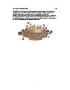
Functions:
Functions of the epithelial tissue include:
- Lining of all body surfaces, both inside and out. Eg, skin and bladder.
- Protection of underlying structures from injury through abrasion or pressure, and from infection.
- Specialised structure, capable of absorption (ointments, water, and electrolytes) secretory (mucus) or excretory (sweat, foreign bodies through cilia).
- Reception of stimuli (through sensory cells and nerve endings).
Structure and Function of the Liver _ P3
The liver is the largest gland in the body. It performs a large number of tasks, which effect the whole body system.
Structure of the Liver
The liver is a specially designed organ, on that it has two sources blood.
The hepatic artery, carrying oxygenated blood from the heart and the portal vein, carrying food substances from the stomach and intestines. About 75% of blood entering the liver is venous blood from the portal vein. Terminal branches of the hepatic portal vein and hepatic artery empty together and mix as they enter minute sinusoids in the liver.
The blood flows through the sinusoids and empties into the central vein of each lobule. These central veins merge into hepatic veins which leave the liver and empty into the vena cava, which then goes into the right heart.
Bile is a secretary and excretory product of hepatocytes in the liver. The hepatocytes secrete bile into the canaliculi, and then flow parallel to the sinusoids, but in the opposite direction that the blood flows.
At the end of the canaliculi, the bile flows into bile ducts, which are lined with epithelial cells. These ducts lie very closely to the terminal branches of the portal vein and hepatic artery, which is known as a portal triad.
Small bile ducts become larger ones eventually forming the common bile duct, this off-loads the bile into the duodenum. When bile is not needed it is diverted into the gallbladder where it is dehydrated and stored until needed. The gallbladder is a muscular organ, which is a membranous sac often pear-shaped. It is found on the under-surface of the right lobe of the liver.
Functions of the liver
The liver has many functions including:
Digestive functions are carried out by bile, which is produced in the liver.
The liver is very important for homeostasis because it directly determines the concentration of many different substances in the blood plasma. These activities are called metabolic or regulatory functions, these include:
- Regulation of blood glucose
- Regulation of lipids
- Regulation of aminoacids
- Regulation of plasma proteins
- Storage of vitamins and minerals
- Production of heat
- Storage of blood
Excretion is the removal from the body of the waste products of the metabolism and of substances surplus to the body’s requirements.
Urea can only be formed in the liver, if ammonia is left to accumulate in the blood it will cause problems.
- Break down of blood cells
Red blood cells are continually being produced in the bone marrow and have a life spa of 129 days. The old blood cells are destroyed all over the body by groups of phagocytic cells. In the liver this is carried out by the kupffer cells.
Detoxification is the removal of toxins and poisons. Bacteria and other pathogens are removed from the blood as it flows by the kupffer cell in the sinusoids. Hepatocytes, which are the major function of the liver, use biochemical reactions to remove toxins.
Kidneys and Osmoregulation M3
Osmoregulation is very important. Tissues do not lose or gain water by osmosis because the concentration of water and salts is the same inside and outside of the cells. The osmotic strength of blood depends upon how much glucose and mineral salts it contains as well as how much water is present.
Dehydration
The hypothalamus detects changes in the amount of water present in the blood. If there is too little water (the blood is too concentrated) it sends messages to the pituitary gland to secrete ADH (Anti-Diuretic Hormone). This hormone has an effect on the kidneys. ADH makes the kidneys reabsorb water from the ultra-filtrate. Higher levels of ADH make the kidneys reabsorb more and more water. This results in the production of very small quantities of very concentrated urine. The result of reabsorbing water is to reduce the concentration of the blood. The hypothalamus detects the change in the blood concentration, sending messages to the pituitary gland to secrete less ADH, this is known as Negative Feedback. The blood then returns to its correct osmotic concentration.
Waterlogging
The hypothalamus detects that there is too much water in the blood. If the blood is too dilute, our cells will absorb water by osmosis and become ‘waterlogged’. Animal cells are in danger of swelling and bursting if they are placed in a solution, which is too dilute. It is very important that the blood does bot become so dilute that the cells become stressed by waterlogging.
When the blood become too dilute, the pituitary gland stops making ADH. The kidneys then stop reabsorbing the water. Large volumes of very dilute urine are formed.
When the concentration of blood starts to rise, the pituitary gland starts to produce ADH again.
Blood Glucose Control M4
It is vital that all animals maintain the concentration of glucose in the body within the normal range.
Some cells, like neurones from the nervous system can only take fuel in the form of glucose, so the concentration of glucose in the blood must be kept over a minimum to keep those specialised cells working.
Glucose levels are kept within the normal limits in several ways:
- The liver stores glucose in the form of glycogen after it receives excess glucose than needed for general circulation, from the portal vein.
When the concentration of glucose in the blood begins to fall, the liver activates the rupture of the glycogen, producing glucose, which is then taken back into the blood for transport to other tissues (glycongenolysis).
The amount of glycogen formed is controlled by a hormone called insulin, this is produced by the pancreas. This hormone acts mainly by prompting the uptake of glucose by the membranes of the liver and muscle cells. In the human liver up to 60g of glucose can be stored in the form of glycogen and 150g in the muscles.
Once this limit has been reached, excess glucose is converted into molecules of fat, which then leave the liver through the vein and are taken up for storage by adipose cells, mostly under the skin.
The process of glycongenolysis is controlled by two hormones, glucagon from the pancreas and adrenalin form the adrenal glands. They are released in response to a fall in blood glucose but adrenalin can also be produced in times of stress so it boosts the availability of glucose quickly.
- When animals have not eaten for several hours, hepatic glycogen reserves become exhausted. The hepatocytes recognise the problem and activate other enzymes to start synthesising glucose out of other substances, like fats or amino acids, if the fasting is prolonged. This process is known as gluconeogenesis, and it is controlled by the hormone glucagon.
The ability of the liver to synthesise glucose this way is greatly important to carnivores, whose diet in the wild has virtually no starch.