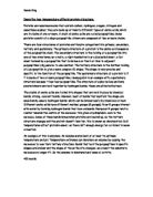
The secondary structure results directly from the primary structure. This involves the polypeptide being changed into mainly two different shapes; alpha helices and beta pleated sheets. These occur because the hydrogen atom of the –NH group of one side of the peptide bond carries an overall positive charge whereas the oxygen in the –COOH carries an overall negative charge. As a result, the hydrogen and oxygen are attracted towards each other weakly, forming hydrogen or H-bonds. This makes the polypeptide chain twist and arrange itself into the alpha helix and beta pleated sheet structure as can be seen in the diagram. The dotted lines show the H-bonds. It requires about 3.6 amino acids to make one ‘turn’ of the alpha helix. Normally, all the alpha helices in a natural protein have a ‘right-hand turn’ that is, the way they turn, like a screw, but some can also have left handed turns.
The tertiary structure of a protein is similar to the secondary structure in that it involves bonds being made. However, the difference is that there are many other types of bond formed between different amino acids so the shape formed by them is much more complicated than the relatively simple helices and pleated sheets. This structure involves four main forces of interaction. Firstly, as in the secondary structure, there are hydrogen bonds which are relatively weak. Then, there are di-sulfide bridges which occur between two sulphur atoms. The only amino acid out of the twenty naturally occurring which can be involved in this type of bond is cysteine. This bond can be seen on the diagram at the top of this page. Another bond is the ionic bond. This occurs between any acidic carboxylic groups (-COOH) and basic amine groups (-NH) that do not contribute to the formation of the peptide bonds. This works by transferring protons from one to another, as a result making a negative carboxylate ion and a positive ammonium ion, which are attracted to each other. Finally, the other type of attraction is the Van der Waal’s forces. They are weak attractions between the non-polar side groups. The structure of a tertiary protein is also affected by the hydrophobic attractions or exclusions; if an atom is hydrophobic, it will face inwards and away from any water-based environment. After all of these bonds and interactions have had an effect, the polypeptide chain looks like this:
Finally, the quaternary structure of a protein makes the polypeptide chain in to a ‘proper’ protein. This involves usually more than one polypeptide chain joining together, along with prosthetic (non-protein) groups to make an overall protein molecule. The way these polypeptides are joined together is similar to the tertiary structure except that instead of the bonds being formed with amino acids of the same polypeptide chain, they are formed with other chains. An example of a prosthetic group is iron, in haemoglobin which is very important in determining its structure and therefore its function. Another example of a prosthetic group is glucose in the protein miraculin. This is named as such because of its ability to make any sour or bitter substance taste sweet. There are two main categories of proteins in this section: fibrous and globular proteins. Fibrous proteins normally have structural functions as they are formed by long, parallel chains which are linked by cross-bridges. An example of this is collagen, used in tendons and keratin, used in hair. Globular proteins carry out metabolic functions and do not have a set structure. Therefore, they are most found as a spherical shape, hence the name globular. Examples of these include haemoglobin and insulin, which will be looked at later. Examples of both types of quaternary structure proteins are shown.
Although all the stages have crucial parts to play in forming the structure of proteins, it is important to note that the secondary, tertiary and quaternary structures all result from the primary structure. A protein can be made if it does not have the secondary or tertiary structure, but in every single protein in existence, there has to be a primary structure. As a result, it can be seen that the primary structure dictates the overall shape of the protein by dictating each of the different stages which in turn make the protein function and work in the correct manner. This idea can be seen more clearly with examples and therefore I have chosen insulin, prothrombin and collagen as three proteins to be looked at in detail. This will show in a practical way how the proteins do their jobs and how this links to the individual and therefore primary structure of the respective protein.
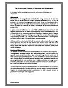
Insulin in humans is made up of two polypeptide chains, with a total of 110 amino acids. The two chains are held together covalently by two di-sulfide bridges. There is also a di-sulfide bridge within the first chain. As can be seen on the diagram, it has a three-dimensional structure, which contains three alpha-helices (brown and blue) and the three di-sulfide bridges (yellow). The protein also has two non-polar surfaces which are involved in Van Der Waal’s forces and so are weakly attracted. The di-sulfide bridges are made up of the amino acid cysteine which has been already mentioned, with the R group of CH2SH. The sulphur contributes to the di-sulfide bridges. Because of the high number of carbon atoms in the centre of many amino acids of insulin, the protein has a hydrophobic core. This is then surrounded by charged hydrophilic amino acids. This, along with the very strong di-sulfide bridge backbone, makes the structure of the protein very stable.
Insulin is very important for organisms because it is central to regulating the carbohydrate and fat metabolism in the body. As it is a cell signalling protein, it is able to send instructions to cells in the liver, muscle and fat to take up glucose from the blood and store it as glycogen or animal starch. It also works the other way round; when blood glucose levels get high, it inhibits the storage of glycogen so the levels of blood glucose can be regulated and brought down again. Because of this, this hormonal protein is essential for organisms like humans.
Prothrombin consists of two activation peptide fragments, a heavy thrombin chain and a light thrombin chain. It contains 622 amino acids which are bonded in a globular structure as can be seen on the diagram. The protein has a very complex globular structure as can be seen on the diagram, with many alpha helices, beta pleated sheets and di-sulfide bridges.
Prothrombin is made by the liver and secreted by the plasma. Its function is very important in the body as it affects blood homeostasis, wound healing and inflammation. However, it can also cause harm to the body; it has been implicated as being a major factor behind a cerebrovascular accident. This is because blood from a ruptured cerebral aneurysm clots around a cerebral artery and releases thrombin. This narrows the artery, resulting in brain stroke or ischemia.
Finally, Collagen is a fibrous protein, composed of three chains wound around each other in a triple helix structure, as can be noticed on the graphic (next page). It is a very large protein as it is made up of more than 1400 amino acids. It has a relatively unusual amino acid composition as almost every third monomer is glycine, maybe because it is small enough to fit easily inside a helix as its R group only consists of a single hydrogen (H) atom. Also, the amino acid proline forms 17% of the protein as a whole. In the tertiary structure, the polypeptide chain is twisted into a second alpha helix. This then joins with three other polypeptide chains very strongly, like fibres wound round a rope which gives collagen its strength.
This strength is very useful throughout the body and without it, many of the functions would have been impossible. It is found almost everywhere in the human body, but mainly in tendons where its long fibrous structure strengthens and enables them to make the bone contract the muscle. It also provides structural benefits to the body as it protects the softer tissues and ensures they remain connected to the skeleton. An idea of the immense influence of collagen can be gained from the fact that the space between nearly every cell in the body is crisscrossed by touch fibrils, formed from a type of collagen. This makes sure that the cells remain stable and as the fibrils act like strong covalent bonds, the structure is held together powerfully.
Collagen is only a polymer; it is actually made up by lots of molecules called tropocollagen. They are approximately 300 nm in length and 1.5nm in diameter. They are formed from three polypeptide strands called alpha polypeptides. As collagen contains millions of strands of tropocollagen and they are all covalently bonded, you begin to get the idea of its strength. Indeed, a single collagen fibre of 1mm diameter has the ability to support a mass of 10kg if not more before it finally breaks. This is why it is so useful in the human body; a massive amount of exertion is needed before it breaks and causes damage.
In conclusion, it can be seen that the primary structure is essential in determining the structure and function of a protein. If the amino acids did not bond in the exact required order, then the protein would fail to work correctly and hence all of the other structures would also fail. Therefore, it is imperative that the primary structure is correct for the rest of the protein to work, even though it is comparatively very simple. Furthermore, it can also be seen that although some proteins do not have secondary or quaternary structures, they must have a primary structure, further highlighting its importance for proteins.