During the 19th century during the industrial revolution, the microscope developed further, as countries started manufacturing fine optical equipment with the help of machinery.
The present light microscope has a maximum resolution of 0.2 µm or 200nm, which is 500 times better than the human eye. It is impossible in theory improve the resolution of the light microscope because of the important limiting factor, which is the wavelength of light (0.4 µm for violet whereas red light has a wavelength of 0.7µm.) Hence only large organelles and cell structure of ekaryotic cells can be seen.
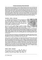
How it works
- Light is produced from either an internal or external light source and passes through the iris diaphragm, a hole of variable size, which controls the amount of light reaching the specimen.
-
The light then passes through the condenser, which focuses the light onto the specimen.
- The slide is held on the stage at 90 degrees to the path of light, which next travels through the specimen.
- The objective lens magnifies the image of the specimen before the light travels through the barrel of the microscope. (3)
Below is a diagram of the light microscope.
The Transmission Electron Microscope
History
The TEM was developed by scientists who wanted to overcome the limiting factor of the light wavelength in light microscopy. The organelles of the cell could not be explored using the light microscope, as this required 10, 000x magnification.
Ernst Ruska from Germany made the breakthrough in 1932 by developing the TEM, with the help of his fellow physicist Max Knoll. For his excellent initiatives and contribution to science he was awarded the Nobel Prize. Ruska was a great physicist, as he knew that electrons could be treated in a similar way to light as they both posses a wave aspect, therefore he could manipulate these waves and concentrate them on the specimen using electromagnetic power.
How it works
High voltage electron beams from a cathode are focused by magnetic lenses on to the specimen. They are then magnified by a series of magnetic lenses until they hit photographic plate or light sensitive sensors - which transfer the image to a computer screen. The image produced is called an electron micrograph (EM).
The transmission electron microscope goes beyond the resolution of the light microscope and has been the main factor and instrument in assembling and gaining all the information about the cells and its organelles.
The electron microscope uses electrons instead of light to produce a magnification of minute details with very high resolving power. Electrons allow the TEM to magnify in higher resolution because of their short wavelengths, in contrast to the light’s longer wavelength. Because of these factors that Ernst Ruska considered when developing the TEM, the electron microscope can therefore magnify up to 500,000 times. Below is evidence of the detail the electron microscope can produce
The Scanning Electron Microscope
This new device was developed from the basis of the transmission electron microscope.
Gerd Binnig and Heinrich Rohrer shared half of a Nobel Prize for their development of the scanning electron microscope.
Differences Between Scanning Electron Microscope and Transmission Electron microscope.
The transmission Electron microscope works by emitting fast moving electron, and then by detecting the electrons, which pass through the specimen by a fluorescent screen, which then produces an image on a visual output port. Also a very important factor is that the specimen for the transmission electron microscope will need to be in ultra thin sections so the electrons can pass through it and an image can be formed.
However in the scanning electron microscope Scanning electron microscopes pass a beam of electrons over the surface of the specimen in the form of a ‘scanning’ beam.
Electrons are reflected off the surface of the specimen as it has been previously coated in heavy metals.
It is these reflected electron beams that are focussed of the fluorescent screen in order to make up the image.
Larger, thicker structures can thus be seen under the scanning electron microscope, as the electrons do not have to pass through the sample in order to form the image.
However the resolution of the scanning electron microscope is lower than that of the transmission electron microscope.
Preparing specimen for viewing
- Fixation: Chemicals preserve material in a life like condition. Does not distort the specimen.
- Dehydration: Water removed from the specimen using ethanol. Particularly important for electron microscopy because water molecules deflect the electron beam which blurs the image.
- Embedding: Supports the tissue in wax or resin so that it can be cut into thin sections.
Sectioning produces very thin slices for mounting. Sections are cut with a microtome or an ulramicrotome to make them either a few micrometers (light microscopy) or nanometres
(electron microscopy) thick.
- Staining: Most biological material is transparent and needs staining to increase the contrast between different structures. Different stains are used for different types of tissues. Methylene blue is often used for animal cells, while iodine in KI solution is used for plant tissues.
- Mounting: Mounting on a slide protects the material so that it is suitable for viewing over a long period. (4)
The Advantages and disadvantages of light and electron microscopes
The light microscope has many advantages, for instance it shows magnification of an object up to 1500 times, which is 500 times better that the human eye. Light microscopes are mobile and portable so you can work anywhere. It is also fairly cheap to purchase and operate, as no other energy is inputted apart from light. An important advantage is that the specimen is not damaged when in preparation, this is important as the specimen may need to be used for biological purposes after the magnification.
However despite these advantages the light microscope has a major disadvantage compared to the electron microscope. This is the fact that the light microscope resolving power cannot be increased because of the limiting factor that is the light wavelength. So therefore theoretically it cannot be improved.
The electron microscope, in huge contrast to the light microscope, it can magnify up to 500,000 times. The short wavelength of electrons enables the electron microscope have high resolving power. However the electron has many disadvantages, as it costs over £1 million, and is also expensive to produce electron beam. It is large and bulky (refer to figure 1.3) and requires a special room. Finally, a vacuum is also needed to carry out the magnification.
Below is a table summarising the above paragraphs.
(5)
Reference:
Figure 1.1:
Figure 1.2: WS- Section A Molecules and Cells 3
Figure 1.3:
Figure 1.4:
(1): (* Helena Curtis and N Sue Barnes 1989 Biology, The Fifth Edition W.H. Freeman, pages 94 – 99.)
(2): ()
(3,4):
(5): Mary Jones, Richard Fosbery and Dennis Taylor (2000) Biology 1, Cambridge, Cambridge University Press, pages 3-10.