As well as the medical disadvantages mentioned above, there are still some other disadvantages of having a MRI scan. There are many claustrophobic people in the world, and being in a MRI scanner is a very disconcerting experience for these people. The machines are also very loud during a scan. The noise sounds like a constant, rapid hammering sound. Patients are given earplugs to soften the sound. The noise is due to the rising electrical current in the gradient magnets being opposed by the main magnetic field. The stronger the main magnetic field, the greater the noise of the machine. MRI machine systems are extremely expensive (the system in Holly House cost £750,000) and therefore the individual scans are just as expensive. (Knee scan is £700 and an abdomen scan is £1000)
The main advantage for MRI scanners is that there are no known biological hazards to humans being exposed to magnetic fields of the strength used in medical imaging today, however most facilities prefer not to scan pregnant women as there has not been much research done in the area of biological defects on a developing foetus, as obviously it is very difficult to conduct such research, as many pregnant women do not want to take this risk. There are many things that MRI is ideal for: -
- Diagnosing multiple sclerosis (MS)
- Diagnosing tumours of the pituitary gland and brain
- Diagnosing infections in the brain, spine or joints
- Visualizing torn ligaments in the wrist, knee and ankle
- Visualizing shoulder injuries
- Diagnosing tendonitis
- Evaluating masses in the soft tissues of the body
- Evaluating bone tumours, cysts and bulging or herniated discs in the spine
-
Diagnosing strokes in their earliest stages
In Holly House, superconducting magnets are used in the machines. The magnet is made from niobium titanium alloy, which gives it superconductivity properties. The wire coiled around the core is surrounded by liquid helium (-269°C). This substance causes the resistance in the wire to drop to zero, this is the critical temperature, and reducing the electrical power needed to operate the system makes the machine much more economical to operate, although cooling the system is still extremely expensive, but the key point is that once the electricity has been introduced into the coils then the current will continue at full strength for years without any more electrical input. These magnets produce a 0.5 tesla to 2 tesla magnetic fields, allowing for much higher quality imaging. This is an extremely large number, and the average MRI scanner has a magnetic field that is 10,000 times larger than the magnetic field of the earth. The entire MRI scanner installation is enclosed in a stainless steel or copper shield known as a Faraday cage which blocks out radio frequency signals from local radio and TV stations that might influence the MRI signals.
How the pictures are taken
The human body is made up of billions of different atoms. The nuclei of these atoms spin around randomly, and the main atom that MRI focuses on is the hydrogen atom. The hydrogen atom is ideal because its nucleus has a single proton and it has a very large magnetic moment. The large moment means that when placed in a magnetic field, the hydrogen atom has a strong tendency to line up with the direction of the field. As the magnet runs along the scanner in the same direction as the body, the magnetic field therefore does the same. The atoms will line up in the direction of either the feet or the head. Many of the atoms cancel out, but as there are so many billions, there are still enough to produce astounding images.
Radio Waves
The MRI machine then applies a radio frequency to the body that is specific only to hydrogen. These waves are directed at the part of the body that is needed to be examined. This pulse causes the protons in the area to absorb the energy that is needed to make them spin once again, but now pointing in a different direction. This part of the process is the ‘resonance’ in Magnetic Resonance Imaging. The radio frequency causes the atoms to spin at a particular frequency and in a particular direction. The frequency of the atoms is called the ‘Larmor frequency’ and can be calculated by the specific tissue being imaged and the strength of the main magnetic field. When the magnetic field is 1 tesla, the frequency is 42.58Mhz, and when the magnetic field is 2 tesla, the frequency of the hydrogen atom is 85.16Mhz. The Larmor frequency can be calculated by knowing the gyromagnetic ratio (also known as the magnetogyric ratio) and the magnetic field, and finding the product.
Gradient Magnets
MRI scanners can ‘slice’ the human body in separate slices only a few millimetres thick. They can do this by using gradient magnets. When gradient magnets turn on and off rapidly, they can compose different slices of the body. They move around inside the main magnet, and thus the main advantage is that a picture can be taken of any particular region in a patient’s body without having to move the patient or the machine. Three gradient magnets are used, so that a patient’s body can be dissected in the sagittal plane, and coronally, as well as axial images. When taking an X-ray, patients have to move after every picture is taken, and CAT scanners cannot slice in the sagittal plane or coronally, so it is immediately obvious why MRI scanners have transformed medical imaging to assist diagnosis. The Gradient Magnets are the cause of the extreme noise in the machines. (Mentioned above).
When the Radio Wave pulse is turned off, the hydrogen atoms begin to return relatively slowly to their original alignment within the magnetic field and release their excess stored energy. When they do this, they give off a signal that the coil around the magnet picks up and sends to a computer system in the radiographer’s office. The system then converts this data into a picture which can then be put onto film, or be sent anywhere in the world via the Internet.
A screenshot of the software used by radiologists. This shows the huge amount of images taken and how they can be manipulated.
Other Uses of the Imaging
If it is necessary, patients can be injected with a special dye that contrasts with normal tissue on a scan. MRI contrast works by altering the magnetic field in the tissue being examined. Normal and abnormal tissue will respond differently to this dye, and therefore different signals will be emitted to the computer system. The severity and the position of the abnormal tissue will then be assessed by the specialist, who can therefore use this information to determine the severity of the condition. The name of the dye that is used is gadolinium. Gadolinium contrasts, have been used for more than 15 years in MRI, and have been extremely well tolerated by patients with a much lower risk of allergic reaction or kidney impairment compared to iodine containing contrasts. New Gadolinium compounds are emerging, such as MS-325, which are designed specifically for imaging of blood vessels. The molecule forms a reversible bond to albumen and stays in the circulation for a longer period of time, allowing high resolution images to be obtained. This offers the potential to safely image the blood vessels of the body with MRI with higher accuracy than currently possible.
A MRI image of the spine.
The Future Of Magnetic Resonance Imaging
MRI is relatively new in medical imaging; it has only been in widespread use for about 20 years whereas x-ray imaging has been around for over 100 years. The future for MRI looks very exciting. Very small scanners are being produced, and there are even scanners developed that can simply be placed on an arm, a foot or a knee. Functional brain mapping which is when someone scans a person's brain while he or she is performing a certain physical task such as squeezing a ball, or looking at a particular type of picture, is helping researchers better understand how the brain works. Research is under way in a few institutions to image the ventilation dynamics of the lungs through the use of hyperpolarized helium gas (helium gas that has had its polarity increased). The development of new, improved ways to image cerebral vascular accidents (strokes) in their earliest stages is ongoing. New MRI scanners are being used with a lower magnetic field (0.1T to 0.5T). These machines do not use a superconducting magnet and therefore the hospitals do not need to worry about cooling these, and therefore the economical implication, however the imaging being recorded has to go through more complex computer systems so that it has the same quality images. New scanners such as PET scanners (Positron Emission Tomography) are coming readily available, and these produce even better images, with the opportunity for images to be constructed in 3-D. however, the NHS is not investing in this technology, there are only 6 of these scanners in Britain whereas Germany and France have over 80 of these each. It looks as if MRI will not be overtaken in the forefront of medical imaging for a long time, especially not in the UK.
A small head scanner.
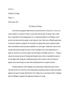
The patient is receiving treatment on his left knee. This is a new type of scanner that is significantly smaller, and therefore has more advantages.
Conclusion
The almost limitless benefits of MRI for most patients far outweigh the few drawbacks. MRI scanning has revolutionised medical imaging, and has helped to treat many patients who could not be treated in any other way. MRI uses physics techniques that have been identified above to overcome problems where CAT scanners could not do so as well. It is an extremely safe method of imaging, and the technology is rapidly advancing, which will only make this method better.
Bibliography
Holly House Hospital MRI Consent Form
neurocog.psy.tufts.edu
www.cksociety.org
hypertextbook.com/facts
‘Economics of the NHS’ BBC2 7th February
‘Medical Physics’ by Martin Hollins, pages 186 - 187