
The advantages and limitations of electron microscopy.
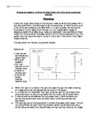
The advantages and limitations of electron microscopy There are two main branches of microscopy that are pertinent to cell biology. These branches arise from the two types of microscope; the light microscope and the electron microscope. The basic principles of light microscopy have been known since circa 17th century, however improvements in lens manufacture in circa 19th century allowed the use of microscopy to be much more practically available and useful. This is increased ability inspired rapid research into both the design of microscopes and the preparation of specimens. However, the light microscope can only magnify objects bigger than 0.2 micrometres; due to its limited resolving powers. This is because it utilises a beam of light. Relatively, light has a long wavelength, this means that when there are two small points close together there is too much refraction and wave front overlap, the eye then only sees one point. This can also be considered in terms of objects "crossing the path" of the wavelength. The smallest wavelength of visible light is 400nm, the diameter of mitochondria is 1000nm, and therefore mitochondria cross the path of the light wave. However ribosomes have a diameter of 22nm, and do not cross the path of the light wave and are therefore not seen by the light microscope. As biologists came to realise these limitations they understood that the

Ray tracing
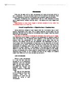
Physics GCSE coursework Ray tracing The objective of this experiment is to find the length of an object and its focal lengths. I will first try to hypothesize where the length points will be. The given results of my hypothesized diagrams will determine the lengths of the focal points. I will then be able to find out where the object image will be situated by following my ray trace diagrams. I believe that I will be able to find the length of the object image if I can find the two focal length points ( 1/v and 1/u displayed in my ray trace diagrams and the focal graph). To achieve this I have decided that I could not get an accurate hypothesis if I did not know this information, so I will carry out a small practical experiment. We found that if the lens is thin, the focal length is longer, and if it is thicker, the focal length is smaller. The focal length for our lens is 10cm. I have created some ray tracing diagrams to show my predicted lengths using the focal length that I found. I have found the longest distance that I can get a clear image is 100cm, the shortest being 15cm. I then carried out an experiment to prove my hypothesis. I used light boxes to create a light source, but this is not extremely accurate as the light rays diverge and are not parallel. I used a screen and a lens to try to find an image. My results for my experiment were as follows. I had three
Physics coursework; Finding the focal length of a lens using a graphical method.
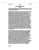
Physics coursework; Finding the focal length of a lens using a graphical method. Planning: Firstly the rough focal length of my lens will need to be found to assist me in my real experiment. A simple way to do this would be, to hold the lens up to a flat white wall opposite a window when it is light outside, by moving the lens closer/ further away from the wall until an upside down image of objects outside the window (e.g. trees,) is produced, I can estimate an focal length for the lens which provides me with the minimum distance of (u), this saves time that would be spent trying to find a point from which I can begin measurements. The equipment will be set up as shown below: Apparatus: * Light source connected to a power pack * Wire grid (object) * 1m ruler (correct to the nearest mm) * a small bi-convex lens * a white 2D screen (approx 100*70 mm) ==> When the light is turned on the light will pass through the mesh creating an image which can be focused by the lens on the screen. ==> It is important to remember that light bulbs will get hot, so precautions should be taken to ensure I am safe from burns during the experiment. ==> It should also be considered that any experiment involving electricity carries risk so due care must be taken when handling any electrical equipment. ==> The light source will be covered with a sheet of grease proof paper, this will
Microscopes. Using electrons instead of light means that the illumination has a much shorter wavelength than light.
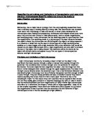
Microscopes Cells can be seen with a light microscope but many structures within a cell - organelles - can only be seen clearly with an electron microscope. That is partly because an electron microscope has a greater magnifying power. However, increasing only magnification has its limits because at some point magnification reveals nothing more - the details only look bigger and vaguer. Magnification is how much bigger a sample appears to be under the microscope than it is in real life. Overall magnification = Objective lens x Eyepiece lens Using electrons instead of light means that the illumination has a much shorter wavelength than light. This is good because minute detail can be detected. We say that an electron microscope has a bigger resolving power than an light microscope Resolution is the ability to distinguish between two points on an image. The resolution of an image is limited by the wavelength of radiation used to view the sample. This is because when objects in the specimen are much smaller than the wavelength of the radiation being used, they do not interrupt the waves, and so are not detected. The wavelength of light is much larger than the wavelength of electrons, so the resolution of the light microscope is a lot lower. The actual resolution is often half the size of the wavelength of radiation used. Thus, for the light microscope the maximum

Electron microscopes.
ELECTRON MICROSCOPY This is the act of using electron microscopes. Electron Microscopes are scientific instruments that use a beam of highly energetic electrons to examine objects on a very fine scale. Electron microscopes can be used to view the topography (surface), the morphology (the shape and size of the particles making up the object) and also the composition (elements and compounds the object is composed of and how many: in case of cell organelles). Electron microscopes were introduced or developed due to the limitation of light microscopes. This is because the resolving power of a microscope depends on the wavelength of the electromagnetic radiation used; because the light microscope uses only the visible part (light) of the electromagnetic spectrum whose shortest wavelength is 400 nanometre (violet light), therefore objects smaller than half of the wavelength (200nm) cannot be viewed using a light microscope. E.g. cell organelle ribosome is 20nm and can never be seen using a light microscope. As electron microscopes uses electrons, which are negatively charged and beams of electrons have a very short wavelength. This type of microscope has a very high magnification and resolution power. They are two major types of electron microscopes the first type originally developed: The Transmission Electron Microscope, which is quite similar to the light electron microscope
Comparing the Light and Electron Microscope
Comparing the Light and Electron Microscope In this essay I am going to be comparing the light and electron microscope, I will look at the advantages and disadvantages of each microscope and then analyse my findings to see if one is better than the other. The light, or optical microscope as it is also known was invented in the 17th century, it has been refined in many ways over the years but it is essentially still the same. The light microscope works by; light rays from a light source beneath the stage are through to glass lenses in series. The two lenses are called the objective lens and the ocular (eyepiece) lens. Depending on their strength these two lenses on their own routinely provide magnifications of up to 400 times. There is a limit to the amount of detail the light microscope can show, this limit is set by the resolving power. The resolving power is the minimum distance by which two points must be separated in order for them to be perceived as two separate points, rather than a single fused image. For the light microscope this distance is approximately 0.2µm. So in theory it might seem possible to magnify an object indefinitely by means of glass lenses in series. This has been put into practice and has only produced a larger and fuzzier picture; so the resolution is not improved and no more detail is visible. The resolution of the light microscope is imposed by

Microscopy. History of the microscope:-
Microscopy Microscopes are tools which allow us to see objects which we cannot see with the naked eye. There are two main types of microscopes used nowadays. These are light microscopes and electron microscopes. During the 16th century the microscope was invented, which was of great assistance to works in medicine and biology. At first, the microscope was basically used recreationally, and was found in the homes of wealthy people. However, not long afterwards, proper uses for the microscope were discovered, and so study of bacteria and diseases began. History of the microscope:- * Circa 1000AD - First vision aid was invented called a reading stone. It was a glass sphere that magnified when laid on top of reading materials. * Circa 1284 - Italian, Salvino D'Armate invented the first wearable eye glasses. * 1590 - Zaccharias Janssen and his son Hans Janssen experimented with multiple lenses in a tube and observed that objects appeared greatly enlarged * 1665 - Robert Hooke noticed some "pores" or "cells" in a sliver of cork looking through a microscope. * 1674 - Anton van Leeuwenhoek built a simple microscope with only one lens to examine blood, yeast, insects and other tiny objects. He invented new methods for grinding and polishing microscope lenses that allowed for curvatures providing magnifications of up to 270 diameters, the best available lenses at that time.
Describe the principles and limitations of transmission and scanning electron microscopes. Specific reference should be made to magnification and resolution
Describe the principles and limitations of transmission and scanning electron microscopes.Specific reference should be made to magnification and resolution Introduction Microscopy has a major role in cytology.From the very beginning researchers have tried to develop ways of looking directly at living cells.This examination has revealed much about the morphology of cells and tissues.In recent years,development in microscopes,dyes,staining and preparatory techniques have helped reveal even more about the structure and function of cells.Microscopes have a certain magnification and resolving power.In any microscope the the resolving power is more important than the magnification.The resolving power of a microscope is the least distance between two objects where the microscope can still distinguish the objects as being separate.It is a measure of detail that can be seen.A microscope with a high resolving power enables us to view images with a high resolution.With a low resolution they would be viewed as one object.Microscopes with a high magnification are only able to increase the size of the object that is being viewed.The resolution will be the same.(ie the object will still lack clarity and appear fuzzy).The two types of microscopes are electron and light microscopes. Principles and Limitations of light microscopy Light microscopes function by focussing a beam of light on

The Principles and Limitations of Electron Microscopy.
The Principles and Limitations of Electron Microscopy. An Electron Microscope is a type of microscope that makes use of a beam of electrons rather than visible light. Which is due to the fact that the wavelength of electrons is much smaller than the wavelength of visible light, an Electron Microscope not only gives a high magnification but it also has high resolution. This means that details can be seen clearly. An Electron Microscope is very similar to a Light Microscope in the ways in which it works but, instead of using glass lenses to focus a beam of light, it uses magnets to focus a beam of electrons. Electrons are very small, so they are scattered if they hit molecules in the air due to this they must travel through a vacuum. Specimens that are to be examined must be cut into very thin sections, and these must be treated so that they can be examined in these conditions. The variety of chemical processes that are carried out in this preparation may change the appearance considerably. Features which have been introduced in this way are known as artefacts and care needs to be taken in interpreting electron micrographs because of the possible presence of artefacts. An examination of a specimen using an Electron Microscope can yield the following information: Topography: The surface features of an object or "how it looks", it's texture; direct relation between these

The Use and Operation of the Light and Electron Microscope
The Use and Operation of the Light and Electron Microscope Light microscopes were first used in the seventeenth century, famously by the scientist Robert Hooke; the man that first named 'cells'. It was not until the nineteenth century however that microscopy became popular. Before this time, the lenses had not been of high enough quality to view images clearly, in the early nineteenth century improvements were made and the identification of cell structures began. Problems with resolution then became apparent; a wave length shorter than light needed to be used in order to improve the clarity of image produced; Electron microscopes were developed as a solution; both Ultraviolet light and X-rays have shorter wave lengths but it was found that these were extremely difficult to focus. Electrons, however, are negatively charged particles; this gives the advantage of being able to focus them easily, by use of electromagnets. These electromagnets act in exactly the same way as a lens would on a Light Microscope. How light microscopes work In a compound light microscope; a light source is located underneath the stage. The light goes through a condenser lens and through the specimen, the resulting light is then passed through two more lenses, both used to magnify the image and focus towards the ocular. The lenses used indicate the resulting magnification of the specimen. Lenses are