
Refraction of Light Lab Report
David Urlanda Physics Lab Report Refraction of Light - Air into Glass Purpose: When light travels through different mediums, it is being refracted. The purpose of this lab is to test Snell's law of refraction. Hypothesis: The angles of refraction that I predicted from the angle of incidences by using Snell's Law are below on the predicted angle Column. To obtain these values I used the index of refraction of crown glass because it is more likely close to the glass (plexiglass) that we are using. Angle of Incidence 0° 0° 20° 30° 40° 50° 60° Predicted angle of refraction 0 6.56° 3.0° 9.2° 25.02° 30.27° 34.74° Variables and Controls: Independent Variable: The angle of the light coming from the ray box or the angle of incidence Dependent Variable: The angle of refraction on the plexiglass. Controls: The mediums where light travels (air and plexiglass). Materials: - ray box - plexiglass - white paper, protractor and pencil Procedure: . I folded the blank paper into four equal parts. Then drew two intersecting lines perpendicular to each other. 2. Using the protractor I drew the angles of incidences or rays measuring 10°, 20°, 30°, 40°, 50° and 60°. 3. Then I drew a semi-circle on the top of the intersection representing the flexi glass and placed the flexi glass over the semi-circle. 4. Plugged in the ray box to a power source
Find the separation between two cones of the same type on the fovea of the eye by using the resolving power of the eye.
Resolving Power Of the Eye Objective: Find the separation between two cones of the same type on the fovea of the eye by using the resolving power of the eye. Introduction: The retina contains two types of light detecting cells: rods and cones. Cones provide the eye's colour sensitivity, rods, though more sensitive than cones do not detect colour. There is an area on the retina with a much higher density of cones called the fovea. When an object is observed its image is focused on the fovea. The fovea is a 0.3mm diameter area containing on rods and very thin densely packed cones. Cones can be divided into three types; one type detects each of red, green and blue light. The green and red cones are concentrated in the fovea centralis. To measure the separation between two cones in the eye we can use the resolving power of the eye, for two objects to be resolved optically the viewer must be able to clearly differentiate two distinct bodies. (Rayleigh's criterion:? = ?/d) Critical case shown where objects are just resolved For two light sources of the same wavelength to be resolved the light must stimulate two cones on either side of one unstimulated cone. Resolving power due to a circular aperture can be calculated by: ? = 1.22? d Where: ? = resolving power of optical instrument ? = Wavelength of light d = diameter of aperture The resolving power of the eye will
An investigation into the workings of the opticians
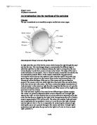
An investigation into the workings of the opticians The eye The eye is considered as an incredibly complex and delicate minor organ. Acknowledgement of image: www.nei.nih.gov/health As light enters the eye it first hits the cornea which focuses the light through the pupil and into the lens. This lens changes shape to accommodate the different angle at which the light hits it so that at whatever distance the light is coming from the lens always focuses it so the focal point is always on the macula. The shape of the lens is controlled by the cilliary muscle. This is a band of muscle around the lens and the two are connected by zonular fibres. As the muscle contracts the ring gets smaller allowing the lens to become more spherical, then when the muscle relaxes the ring grows larger again and pulls on the zonular fibres so the lens becomes flatter. The vitreous gel retains the shape of the eye so all the cornea and lens can function properly. The macula is the area which light hits at the back of the eye and the fovea is the small yellow dot in the centre. The fovea contains the highest concentration of 'rods and cones' in the macula so light from the image that hits this area will give the clearest and sharpest picture. Light that hits the rest of the macula will be slightly less clear but will still be in focus The 'rods and cones' spoken of are actually two different types

Microscopes. Using electrons instead of light means that the illumination has a much shorter wavelength than light.
Microscopes Cells can be seen with a light microscope but many structures within a cell - organelles - can only be seen clearly with an electron microscope. That is partly because an electron microscope has a greater magnifying power. However, increasing only magnification has its limits because at some point magnification reveals nothing more - the details only look bigger and vaguer. Magnification is how much bigger a sample appears to be under the microscope than it is in real life. Overall magnification = Objective lens x Eyepiece lens Using electrons instead of light means that the illumination has a much shorter wavelength than light. This is good because minute detail can be detected. We say that an electron microscope has a bigger resolving power than an light microscope Resolution is the ability to distinguish between two points on an image. The resolution of an image is limited by the wavelength of radiation used to view the sample. This is because when objects in the specimen are much smaller than the wavelength of the radiation being used, they do not interrupt the waves, and so are not detected. The wavelength of light is much larger than the wavelength of electrons, so the resolution of the light microscope is a lot lower. The actual resolution is often half the size of the wavelength of radiation used. Thus, for the light microscope the maximum

Aim To determine the refractive index of a material and the speed of light in order to calculate the expected critical angle at which total internal reflection occurs
Practical Experiment 3: Refraction and Reflection of Light Aim To determine the refractive index of a material and the speed of light in order to calculate the expected critical angle at which total internal reflection occurs. Hypothesis As the sin of the angle of incidence increase, the sin of the angle of refraction also increases Theory There is a strong relationship between the angle of incidence and angle of refraction of lights. Light can pass through different materials, assuming that it is transparent. It can also pass through two different isotropic materials such as air to glass. When light passes from one isotropic material with a high refraction index to another isotropic material with a lower refraction index, there is an angle where light passing through gets reflected and refracted (Young 2011). Snell brought up a law, which determines the angle at which light bends according to the initial angle and also the refraction index of materials. Where he derived the formula; where = refractive index of material a = refractive index of material b = sin of angle of incidence = sin of angle of refraction (Young 2011) However light may not be refracted all the time, as there is a point where the light will not pass the second material, this is when light gets reflected at the surface. This angle is known as the angle of refraction. Equipment: Light Box

The history, development and use of the light and electron microscope
The history, development and use of the light and electron microscope History of light microscope Observing objects in detail greater than the naked eye was very interesting to people at early stage. This led to the construction, in the 16th century, of a magnifier composed of a single convex lens, and this, in turn, led to the eventual development of the microscope. The most famous early pioneers in the history of the microscope are Digges of England and Hans and Zcharias Janssen of Holland. But it was Antony van Leeuwenhoek who became the first man to make and use a real microscope. Leeuwenhoek ground and polished a small glass ball into a lens with a magnification of 270X, and used this lens to make the world's first optical microscope. Because it had only one lens, Leeuwenhoek's microscope is now referred to as a single-lens microscope. Its convex glass lens was attached to a metal holder and was focused using screws. The light microscope system was invented in the seventeenth century. This type of microscope incorporates more than one lens so that the image magnified by one lens can be further magnified by another. Today, the term "microscope" is generally used to refer to this type of compound microscope. Since its invention, the light microscope has made tremendous progress and help figure out many biological molecules. Using a light microscope that he had

Electron Microscopes
Biology - Electron Microscopes Electron Microscopy is the use of Electron Microscopes. Electron Microscopes have a very high resolving power and a high magnification, and thus are used to view small objects with greater magnification and detail than a light microscope. There are two different types of electron microscopes, each with different abilities and limitations. This essay will analyse and discuss the functions and limitations of each different type individually, and then conclude with comparisons. Transmission Electron Microscope This type of electron microscope allows the user to view a 2d image of a cross section of a sample. It has a maximum resolution of 1 nm and a maximum magnification of 250,000 x. The good resolution is due to the short wavelengths of electrons, which is 0.005 nm. In a TEM the electrons are fired from an electron gun, which is part of the cathode, and are drawn through the microscope by the anode. The electrons pass through the specimen, which must be very thin, and prepared in a certain way for exactly that reason. The specimen preparation process will be explained in detail later in the essay. There are three electromagnetic lenses in a TEM. Electromagnetic lenses are used because firstly electron beams cannot pass through glass, and secondly, as electrons are charged particles, they are affected by a magnetic field. The first is called

Electron Microscopy.
Electron Microscopy. Electron microscopy is a method of imaging that uses an electron microscope to enlarge small specimens by a greater magnification and resolution than conventional light microscopes. The photographs produced of specimens viewed with an electron microscope are call electronmicrographs. Magnification is the increase in apparent size of the specimen and resolution (also called resolving power) is the ability of the microscope to distinguish and produce separate images of closely placed objects. These two primary properties of electron microscopes make them extremely useful in the analysis and study of specimens. The obvious difference between electron microscopes and light microscopes is the medium through which each operates. Light microscopes work by using photons to produce an image whereas electron microscopes use electrons to produce an image. The magnifying power of a light microscope is limited by the wavelength of visible light so electrons are used instead because they have a much smaller wavelength so can therefore resolve much smaller structures. The resolving power of a microscope depends on the wavelength of the electromagnetic radiation used. However, the benefits gained by using an electron microscope also bring specific problems that have to be tackled. Electron beams cannot pass through glass because electrons are physical matter.
Electron Microscopy.
Electron Microscopy An electron microscope can only show dead structures, but will show cell ultrastructure including the fine structure of the cell organelles. When using an electron microscope, the specimen is illuminated in an electron microscope by an electron beam. The electron beam is focused using electromagnets arranged around the path of the electron beam. These electrons then produce an image when focused onto a fluorescent screen. This image is formed from electrons, which have been emitted or reflected from the surface of a complete specimen. There are two different types of electron microscopes: a transmission electron microscope and a scanning electron microscope. The electrons pass through or past a thin section of the specimen in a TEM on their way to the fluorescent screen or photographic film. In SEMs the electrons are reflected off the prepared surface of the specimen. SEMs are very useful for detailed study of surfaces. They both have the same wavelength of 0.005nm, they both magnify non-living tissue specimens and they both produce a monochrome image. There are also some differences between them - the transmission electron microscope (TEM) requires a small copper grid as a support, whereas the scanning electron microscope (SEM) requires a small metal disk. Also, the TEM has a maximum magnification of 250,000 times, and the SEM has a maximum magnification
Electron microscopy.
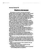
Veronica Ouyang 12C Electron microscopy The development of the electron microscope (EM) has had a significant impact on science research. Invented in the 1930s, the present day version of the EM can magnify up to 500,000 times and has a resolution of about 1nm. In contrast, the light microscope can magnify an object by a maximum of 1500 times and the resolving power is 200nm. That means organelles, which are only blurred images when viewed with a light microscope, can now be studied in great details. Many new structures therefore have been discovered using the EM. Instead of using light, the EM uses a beam of electrons to resolve objects. The beam, which is produced by a heated filament, can be bent and focused by electromagnetic lenses, in the same way the glass lenses are used in a light microscope. The image is projected into a cathode ray tube, rather than the retina of the eye, to make it visible to the operator. When suitable sections are found, they are photographed to give a permanent record-an electron micrograph. The reason why the EM has higher resolving power is that the wavelength of the light used in light microscopes is around 500-650nm. This is much longer than which of the electrons is. That means two objects separated by less than 200nm will appear as one object for the light can not pass though, whereas electrons can. The EM includes two main types-the