
An Essay about Microscopes
Microscopes The word Microscope it given to the tool used to view object that are too small to be seen with the naked eye. During the 1st century AD, glass had been invented and the Romans were looking through the glass. They experimented with different shapes of clear glass and one of their samples was thick in the middle and thin on the edges. They discovered that if you held one of these pieces of glass over an object, the object would look larger. Before microscopes as we know where invented, what was considered as a microscope was just really a Magnifying Lens, early biologist used them to study tiny insects such as Fleas, thus the viewer was called a Flea Lens. Two Dutch spectacle makers in the 1590's (Zaccharias Janssen and his father Hans) experimented with lenses and realised that if several lenses were put into a tube and the object is viewed it appears larger than viewed by any magnifying lens, this was the invention of the Compound Microscope. Anthony Leeuwenhoek of Holland who worked in a dry goods store had a great interest in lens and began making some of his own. By grinding and polishing, he was able to make small lenses with great curves. His rounder lenses produced greater magnification. Anthony Leeuwenhoek's new microscope got him interested in science and with his new improved microscope was able to see things that no man had ever seen before. He saw
Physics coursework; Finding the focal length of a lens using a graphical method.
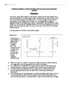
Physics coursework; Finding the focal length of a lens using a graphical method. Planning: Firstly the rough focal length of my lens will need to be found to assist me in my real experiment. A simple way to do this would be, to hold the lens up to a flat white wall opposite a window when it is light outside, by moving the lens closer/ further away from the wall until an upside down image of objects outside the window (e.g. trees,) is produced, I can estimate an focal length for the lens which provides me with the minimum distance of (u), this saves time that would be spent trying to find a point from which I can begin measurements. The equipment will be set up as shown below: Apparatus: * Light source connected to a power pack * Wire grid (object) * 1m ruler (correct to the nearest mm) * a small bi-convex lens * a white 2D screen (approx 100*70 mm) ==> When the light is turned on the light will pass through the mesh creating an image which can be focused by the lens on the screen. ==> It is important to remember that light bulbs will get hot, so precautions should be taken to ensure I am safe from burns during the experiment. ==> It should also be considered that any experiment involving electricity carries risk so due care must be taken when handling any electrical equipment. ==> The light source will be covered with a sheet of grease proof paper, this will
Electron microscopes allow the viewer to see much smaller objects than the original light microscopes. The wavelength of electrons is thousands of times smaller than the wavelength of light
Electron Microscopy Electron microscopes allow the viewer to see much smaller objects than the original light microscopes. The wavelength of electrons is thousands of times smaller than the wavelength of light, with a ratio of around 0.01nm : 500nm, electron wavelength : light wavelength. This overcomes some of the problems faced with light microscopes, such as the depth of focus that can be obtained, the obstruction of objects and the blurred images that often occur due to the wavelengths of light. Electron microscopes can therefore show more detail then light microscopes and can be used to study much smaller objects in finer detail. As electrons cannot be seen as light can, the image is shown on a screen when the electrons are detected within the microscope. A stream of electrons is formed at a hot cathode, or 'electron gun' inside the microscope, which is then 'fired' at the object being viewed using positive electrical charges. Metal apertures and magnetic lenses are used to confine the stream into a thinner, more focused beam. The beam is then directed onto the sample by magnetic lenses. The electrons are detected by a fluorescent screen or photographic plate at the front of the microscope, causing the screen to glow and producing an image of the subject. Due to the finite energy of the electrons, care must be taken in the preparation of the specimen for viewing, and
How to use a Light Microscope
How to use a Light Microscope Introduction: In this coursework I will be using a simple light microscope to look at the letter 'e' which will be cut out from a past newspaper. I will be trying to find out how a microscope works, and what I see through the eyepiece when looking at the letter 'e' and what changes occur when changing the focus and slide. Before I begin the experiment I will be introducing exactly what a microscope is and how it is used. Microscopes are instruments that magnify tiny objects or reveal fine details on larger objects, they reveal to us things that are invisible to our eyes such as cells. The two commonly know microscopes are 'Electron Microscopes' and 'Optical Microscopes', electron microscopes use a beam of electrons controlled by magnetic fields instead of light and electron microscopes are very powerful and can show details 1000 times larger than optical microscopes but the specimen must be dried out and sliced very thinly. In an optical microscope, light shining through an object is bent as it passes through a lens and this will make the object appear bigger than it is and by adding a second lens the magnification will then increase. Optical microscopes with several lenses are called compound microscopes and this is what I will be using in the experiment. Equipment/Apparatus: * Cover Slip * Slide * Small Strip of Newspaper with the
Microscopes. Using electrons instead of light means that the illumination has a much shorter wavelength than light.
Microscopes Cells can be seen with a light microscope but many structures within a cell - organelles - can only be seen clearly with an electron microscope. That is partly because an electron microscope has a greater magnifying power. However, increasing only magnification has its limits because at some point magnification reveals nothing more - the details only look bigger and vaguer. Magnification is how much bigger a sample appears to be under the microscope than it is in real life. Overall magnification = Objective lens x Eyepiece lens Using electrons instead of light means that the illumination has a much shorter wavelength than light. This is good because minute detail can be detected. We say that an electron microscope has a bigger resolving power than an light microscope Resolution is the ability to distinguish between two points on an image. The resolution of an image is limited by the wavelength of radiation used to view the sample. This is because when objects in the specimen are much smaller than the wavelength of the radiation being used, they do not interrupt the waves, and so are not detected. The wavelength of light is much larger than the wavelength of electrons, so the resolution of the light microscope is a lot lower. The actual resolution is often half the size of the wavelength of radiation used. Thus, for the light microscope the maximum

The history, development and use of the light and electron microscope
The history, development and use of the light and electron microscope History of light microscope Observing objects in detail greater than the naked eye was very interesting to people at early stage. This led to the construction, in the 16th century, of a magnifier composed of a single convex lens, and this, in turn, led to the eventual development of the microscope. The most famous early pioneers in the history of the microscope are Digges of England and Hans and Zcharias Janssen of Holland. But it was Antony van Leeuwenhoek who became the first man to make and use a real microscope. Leeuwenhoek ground and polished a small glass ball into a lens with a magnification of 270X, and used this lens to make the world's first optical microscope. Because it had only one lens, Leeuwenhoek's microscope is now referred to as a single-lens microscope. Its convex glass lens was attached to a metal holder and was focused using screws. The light microscope system was invented in the seventeenth century. This type of microscope incorporates more than one lens so that the image magnified by one lens can be further magnified by another. Today, the term "microscope" is generally used to refer to this type of compound microscope. Since its invention, the light microscope has made tremendous progress and help figure out many biological molecules. Using a light microscope that he had