
How the transmission and scanning electron microscopes work?
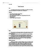
How the transmission and scanning electron microscopes? Introduction Electron microscopy is an important tool in biology in general and cellular biology in particular. The two most important types of electron microscopes are the Transmission Electron Microscope (T.E.M.) and the Scanning Electron Microscope. (S.E.M.) these two types of microscopes are both used in medical, biological and material research, and both have advantages and disadvantages over each other, which I will be mentioning in this essay. The transmission electron microscopes (T.E.M.) The preparation for the T.E.M. Firstly the specimen is fixed, in other word it is preserved in as close to its living state as possible. The specimen then has to be dehydrated with an alcohol (e.g. ethanol). This is because the T.E.M. operates under a high vacuum and it is therefore no water should be to enter the microscope, to prevent this to happen the specimen is placed in liquid resin (similar to araldite glue). After a few days the specimen is heat-polymerised, which produces a firm specimen block, then very thin sections are cut using an ultramicrotome (a special cutting tool). These sections are then stained with solutions of heavy metals to produce a contrast in the observed image by causing diffraction of the electron beams. How the T.E.M. works? The T.E.M. works on the same basic principles as a light
To investigate and demonstrate how the different wavelengths of red and blue light differ by finding their focal lengths using a converging lens.
Physics Coursework Aim To investigate and demonstrate how the different wavelengths of red and blue light differ by finding their focal lengths using a converging lens. Apparatus * Red and Blue LED's (light emitting diodes) * Wires to connect apparatus together * Power supply and mains access to control voltage supplied to the LED's * Ruler in cm and mm * Converging lens * Blocks to adjust height of components Safety This experiment is relatively safe and there are few hazards. However I will be aware throughout the experiment of the electrical components thus minimising any risk of electric shock. Although LED's them selves do not get sufficiently hot enough to burn skin the wires may get quite hot if the current passing through them is high enough. If I conduct the experiment with a high voltage not only may the LED's fuse, the brightness of them may harm my eyes if they are looked at continuously. To reduce this effect I shall have a small voltage but with a high enough brightness within the LED to obtain accurate results. Chromatic Aberration Different wavelengths are refracted by different amounts. The refractive index is different for different colours. This leads to an effect called chromatic aberration. A simple lens has different focal lengths at different wavelengths (Colours). This is because the different colours have been refracted through the

Identification of purpose of physics.
Greenwich Royal Observatory Identification of purpose of physics From my visit to the Greenwich Royal Observatory, the first aspect of physics that I will be showing an understanding for is the time keeping due to the earth's movements. Time keeping is on of the most important and what make Greenwich the most famous place for the "creation" of measuring time efficiently. Everyone needs the time and that's why so many years were put into inventing the "perfect" clock. The physics behind the clock is to get an exact measurement of 1 second. This has been achieved by pendulums, weights and now by computer. Time has been completely based on the earth's movement. Early on time was just measured by the light of day but then this all changed due to the stars. In the 1700's people started to measure the earths spin by using the starts. At night the looked up and pinpointed a star. The following nigh they would wait for that start to appear in the same place. The time between this was 24 hours. This was a great result due to the fact that there were 12 months in a year and this could easily let the time "integrate" with this number. However they shortly found out that seconds were being gained and lost due to the earth spinning round the sun. This means that they need some way in keeping the time correct after years. This is here the atomic watches came in. These

Electron Microscopy.
Electron Microscopy. Electron microscopy is a method of imaging that uses an electron microscope to enlarge small specimens by a greater magnification and resolution than conventional light microscopes. The photographs produced of specimens viewed with an electron microscope are call electronmicrographs. Magnification is the increase in apparent size of the specimen and resolution (also called resolving power) is the ability of the microscope to distinguish and produce separate images of closely placed objects. These two primary properties of electron microscopes make them extremely useful in the analysis and study of specimens. The obvious difference between electron microscopes and light microscopes is the medium through which each operates. Light microscopes work by using photons to produce an image whereas electron microscopes use electrons to produce an image. The magnifying power of a light microscope is limited by the wavelength of visible light so electrons are used instead because they have a much smaller wavelength so can therefore resolve much smaller structures. The resolving power of a microscope depends on the wavelength of the electromagnetic radiation used. However, the benefits gained by using an electron microscope also bring specific problems that have to be tackled. Electron beams cannot pass through glass because electrons are physical matter.
Investigation of the chromatic aberration of a converging lens.
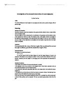
Investigation of the chromatic aberration of a converging lens By Wen Yan Gao Aim To find the difference in focal length of a converging lens when used to produce images with red and blue light. Planning Background information Chromatic aberration arises from dispersion- the property that the refractive index of glass differs with wavelength of light. The focal length of a lens is determined by a combination of its geometry and the refractive index of the material from which it is made. The refractive index varies slightly with the wavelength of the light that is being transmitted. This means that the focal length of a lens will vary for different colours of light. For blue light (short wavelengths), the focal length is larger than that of red light (long wavelengths). Prediction It was predicted that the values of the focal length of the lens obtained from red and blue light are different, given that the standard focal length is 10cm. Proposed Method: The method is to use the lens formula. 1/f = 1/u +1/v To use this formula both the object distance (u) and the image distance (v) need to be measured. This method is not as simple as method 1 above, but the result obtained will be more accurate. Therefore, the focal length of a converging lens in this experiment was determined by method 2. Detailed Procedures: All the lights were switched off to ensure the

Determine the effect changing the object to the lens distance has on the distance at which the image is displayed.
Lenses Aim: To determine the effect changing the object to the lens distance has on the distance at which the image is displayed Prediction: To make my predictions I decided to use a quantitive prediction method, the equation for this is: 1 = 1 + 1 focal length (cm) Object-lens distance Lens-image distance To use this method I will have to know the focal length of the lens. To do this we did a preliminary experiment where I put a lens opposite a window with a piece of paper behind. I moved the paper back and forth until the image of the outside became clear on the paper. The distance from the lens to the card, when the image is in focus gives the focal length. Having established the focal range of the lens was 10.5, and that at this distance the image would not become clear for infinity. Here is our predictions using the equation explained earlier: 1 = 1 = 1 - 1 = 11 - 10 - 1 = V=110 V 10 11 110 110 110 2 = 1 = 1 - 1 = 6 - 5 = 1 = V = 60 V 10 12 60 60 60 3 = 1 = 1 - 1 = 13 - 10 = 3 = V = 43.3 V 10 13 130 130 130 4 = 1 = 1 - 1 = 7 - 5 = 2 = V = 35 V 10 14 70 70 70 5 = 1 = 1 - 1 = 3 - 2 = 1 = V = 30 V 10 15 30 30 30 Prediction shows that the

What are Light Microscopes?
Pass1 What are Light Microscopes? What does the word microscope mean: The first part of the word "Micro" means tiny. The "scope" part means to look at or view. Microscopes are tools that are used too enlarge images of small objects so that they can be studied. A light microscope is an instrument made up of two lens they are eyepiece lens and the object lens combined they produce a much greater magnification that what is possible with just one single lens. The microscope also has a variety of knobs to focus the picture seen thought the microscope. The light microscope is also known as the compound microscope this is because it uses more than one lens. The light microscope uses visible light to detect small objects; the microscope consists of an optical instrument that magnifies the image of an object. It is probably the most used research tool in biology. The total magnification is calculated by multiplying the magnification of the two lenses inside the microscope. Images looked at under the light microscope are reversed and inverted. Functions Of The Components Seen Under A Light Microscope Cytoplasm: is a partly fluid material, which can flow slowly and in which many other substances are suspended such as large fat and protein molecules. Many of the chemical reactions take place in the cytoplasm, which will provide the cell with energy and allow it to build up

The electron microscope and biological advancement.
Toby Nicholson The electron microscope and biological advancement What is an electron microscope? An electron microscope does a similar job to an ordinary optical microscope that many people will have used before - it makes objects appear bigger. This is called magnification and lets one see details of very small objects, such as cell structures, which cannot be seen with the naked eye. In a normal optical microscope this is achieved by using higher-power lens that magnify the image more times. However, there comes a point at which magnification of an optical microscope does not show more detail. This is to do with the physical nature of light. Light is made up of particles (called photons) that travel in a wave. We can only see that two objects are separate if light can pass between them. Light can pass between the letters on this page so you can see them. When objects are very close together light cannot pass through them and they appear as one object. You have experienced this phenomenon when a car heads towards you at night, at first it appears as one light but as it approaches it's head lights split into two headlights. This ability to distinguish between objects is called resolution. The size of wavelength of light limits its resolution to about 2?m, the size of large bacteria. Electrons have a much smaller wavelength than light. This means that they can pas between
Refraction of Light Lab Report
David Urlanda Physics Lab Report Refraction of Light - Air into Glass Purpose: When light travels through different mediums, it is being refracted. The purpose of this lab is to test Snell's law of refraction. Hypothesis: The angles of refraction that I predicted from the angle of incidences by using Snell's Law are below on the predicted angle Column. To obtain these values I used the index of refraction of crown glass because it is more likely close to the glass (plexiglass) that we are using. Angle of Incidence 0° 0° 20° 30° 40° 50° 60° Predicted angle of refraction 0 6.56° 3.0° 9.2° 25.02° 30.27° 34.74° Variables and Controls: Independent Variable: The angle of the light coming from the ray box or the angle of incidence Dependent Variable: The angle of refraction on the plexiglass. Controls: The mediums where light travels (air and plexiglass). Materials: - ray box - plexiglass - white paper, protractor and pencil Procedure: . I folded the blank paper into four equal parts. Then drew two intersecting lines perpendicular to each other. 2. Using the protractor I drew the angles of incidences or rays measuring 10°, 20°, 30°, 40°, 50° and 60°. 3. Then I drew a semi-circle on the top of the intersection representing the flexi glass and placed the flexi glass over the semi-circle. 4. Plugged in the ray box to a power source

An Essay about Microscopes
Microscopes The word Microscope it given to the tool used to view object that are too small to be seen with the naked eye. During the 1st century AD, glass had been invented and the Romans were looking through the glass. They experimented with different shapes of clear glass and one of their samples was thick in the middle and thin on the edges. They discovered that if you held one of these pieces of glass over an object, the object would look larger. Before microscopes as we know where invented, what was considered as a microscope was just really a Magnifying Lens, early biologist used them to study tiny insects such as Fleas, thus the viewer was called a Flea Lens. Two Dutch spectacle makers in the 1590's (Zaccharias Janssen and his father Hans) experimented with lenses and realised that if several lenses were put into a tube and the object is viewed it appears larger than viewed by any magnifying lens, this was the invention of the Compound Microscope. Anthony Leeuwenhoek of Holland who worked in a dry goods store had a great interest in lens and began making some of his own. By grinding and polishing, he was able to make small lenses with great curves. His rounder lenses produced greater magnification. Anthony Leeuwenhoek's new microscope got him interested in science and with his new improved microscope was able to see things that no man had ever seen before. He saw