Above: A phospholipid. (image: Brussel, T V. 2010).
A. Hydrophilic head
B. Hydrophobic tail
As pictured in the diagram below, this causes all of the heads to line up next to each other giving the surface of the cell membrane a charge whilst all of the tails face the inside of the membrane where there is no charge (Biology Arizona, 2004). This polar surface on the top and bottom of the double layer arrangement allows the cell to be exposed to water on both sides (ibid).
Above: The phospholipid bilayer. (Image: Hill, M. 2009).
The lipid bilayer is a highly impermeable structure. Only water and small uncharged molecules such as carbon dioxide and oxygen can cross the plasma membrane with relative ease (S-cool, n.d.). This means that larger charged molecules and small polar molecules cannot cross the bilayer and must pass through specific channels to enter the cell. This is where proteins come in; the proteins can be found within the membrane and they regulate the transport of what substances can get into the cell and what can’t (ibid). They act as pumps or channels which open to allow certain substances to come into the cell and close to prevent unwanted particles entering.. This allows bigger charged substances such as glucose to move across (ibid).
The lipid bilayer is a fluid and flexible structure. This arrangement allows the structures within the bilayer to be mobile and flow around the cell throughout the plasma membrane (Vision Learning, n.d.). This fluidity is important as it influences membrane transport (ibid). Fluidity is dependent on both the specific structure of the fatty acid chains and temperature (ibid). Structurally, the lipid bilayer is irregular: the lipid and protein arrangement in each of the two layers is different (Shuster, 2003).
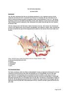
The Cell Surface & Membrane Proteins
The protein and lipid cell membrane is covered with a layer of carbohydrate on the outer surface. This layer is called a cell coat or glycocalyx and provides the cell with protection against damage (S-cool, n.d.). The polysaccharide chains that appear on the external surface of the plasma membrane are involved in cellular communication and help the cell to identify and communicate with other cells in close proximity (ibid). These antibodies are known as glycoproteins (attached to proteins) and glycolipids (attached to phosphate heads) (Vision Learning, n.d.). They are especially important in the immune system as it helps the cell to identify foreign substances and take the appropriate action to eliminate any alien or harmful substances (ibid).
The cell membrane also contains and number of special membrane proteins imbedded in the phospholipid bilayer which perform certain functions. Membrane proteins are important as they allow cells to communicate with their environments, determine whether the immune system recognises the cell as foreign or not, aid the transport of molecules across the membrane, and control cell adhesion to form tissues (Biology Arizona, 2004). These proteins also control development of plants and animals and important metabolic processes such as energy production and transmission, and photosynthesis (ibid). Integral proteins are usually trans membrane proteins, they can extend through the lipid bilayer so that one end contacts the inside of the cell and the other touches the exterior. Many integral proteins are glycoproteins, which have an attached carbohydrate chain (Shuster,2003). The stretch of the integral protein within the bilayer is hydrophobic and made up of non-polar amino acids whilst the exposed ends of the integral protein are hydrophilic (ibid). As a result of their structure, transmembrane proteins are the only class of proteins that can perform functions both inside and outside of the cell. In addition to integral proteins there are also less mobile peripheral proteins which attach to the outside of the lipid bilayer (ibid). They are involved in transferring genetic material from one cell to another and are a mechanism which the cell can use to determine what is in its environment (ibid).
Above: Membrane proteins. (Image: Gwen V. 1995).
A. Carbohydrate group of glycoprotein
B. Peripheral protein
C. Carbohydrate group of protein
D. Carbohydrate group of glycolipid
E. Amino acid chain
F. Transmembrane proteins
Cholesterol
One of the most important tasks that cholesterol performs in the body is in cells. Present in animal cell membranes and absent in bacteria and most plants, cholesterol makes the bilayer stronger, more flexible, less fluid, and less permeable to water-soluble substances such as ions and monosaccharide (Vision Learning, n.d.). This helps stabilise the plasma membrane by stopping it from becoming fluid and splitting apart. It also helps to connect phospholipids together and prevents them from drifting apart in warm temperatures or sticking together during cooler temperatures (ibid). Receptors on the outside of the cell seize cholesterol from the bloodstream as needed to keep the cell functioning correctly (ibid).
Above: Cholesterol. (Image: Gwen V. 1995).
- Cholesterol
Bibliography
Websites:
Biology Arizona. (August 2004). Cell Membrane Tutorial. Retrieved 31st May, 2012, from
Brussel, T V. (2010). A phospholipid. [Online image]. Available at:
(Accessed: 31st May 2012).
Gwen V. (1995). Membrane proteins. [Online image]. Available at:
http://www.cytochemistry.net/cell-biology/membrane_intro.htm (Accessed: 31st May 2012)
Gwen V. (1995). Cholesterol. [Online image]. Available at:
http://www.cytochemistry.net/cell-biology/membrane_intro.htm (Accessed: 31st May 2012)
Hill, M. (2009). The phospholipid bilayer. [Online image]. Available at:
(Accessed: 31st May 2012).
Pietzsch, J, (2004). The fluid mosaic model of membrane structure. [Online image]. Available at:
(Accessed: 31st May 2012).
S-cool. (n.d.). Cell Membrane. Retrieved 31st May, 2012, from
S-cool. (n.d.). Movement. Retrieved 31st May, 2012, from
Shuster, C. (2003), Cells: Discovery and Basic Structure, Retrieved 31st May, 2012, from http://www.visionlearning.com/library/module_viewer.php?mid=64
Vision Learning. (n.d.). Cytoskeleton - the movers and shapers in the cell. Retrieved 31st
May, 2012, from
The Open University. (2002). Cell membrane. Retrieved 31st May, 2012, from
Other:
‘Active Transport’. (A4 Bio-factsheet hand-out from Biology lesson 16th May 2012).
Earley, B. 2012, Class notes, Cell Membrane Introduction, Biology lesson 25th April 2012.
Earley, B. 2012, Class notes, Cell Membrane drawing, Biology lesson 25th April 2012.