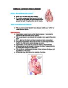
The heart is divided into a right and a left side both separated by a thin wall. Each side is further divided, making four chambers. The two upper chambers of the heart, called the atrium, collects blood and the lower two chambers both called the ventricle, have thicker walls which allows blood to be pumped from here, around the body. Between each atrium and ventricle is a non-return valve, which allows the blood to flow in one direction only. (See diagram b)
The blood is pumped around the body by rhythmic contractions, these are the beats of the heart muscle. Each beat has three phases, the Atrial Systole is when blood is squeezed out of the atria, (the top chamber to the bottom chamber). The Ventricle Systole is when blood is forced to the Aorta, to the rest of the body, and the pulmonary artery, which goes to the lungs, and then the arterial diastole, which means the heart, relaxes and allows blood to fill into the atria. The ventricles then relax this is called Ventricular Diastole.
Muscle cells within the heart are joined together in a branching network, this is so each cell is in intimate contact with at least two of its neighbours. Due to the chemical process, which occurs within it, each cell builds up an electrical charge across the membrane that surrounds it. The outside of the cell becomes positively charged and the inside of the cell, negatively charged. Immediately before contraction, ‘the cell membrane leaks and the ‘voltage’, (Barnard.C, The Body Machine, 1978, page 80) difference between the two sides is momentarily dissipated by a transient short circuit’. This is known as depolarisation and is followed by contraction of the cell. After the contraction, the leaky cell membrane is repaired and the voltage across it is restored by repolarisation. The entire circle of depolarisation, contraction, then repolarisation is repeated continuously. The heartbeat is controlled by a pacemaker or initiating centre for this electrical impulse is known as the sinus node. This is a specialised group of cells, which is situated in the right atrium close to the point of entry of the great vein draining the upper half of the body. The sinus node initiates each wave of depolarisation, which then passes through the muscle cells of the atria to a second group of specialised cells, which is situated at the junction of the atria and ventricles. This is known as the atrioventricular node. From here the impulses pass down two bundles of specialised heart cells running along either side of the wall separating both the left and right ventricles. These bundles distribute the impulses to the muscle cells of the right and left ventricles.
The journey of the blood starts from the left side of the heart through the large artery, the aorta. At this stage the blood is rich in oxygen, food broken down into the microscopically small components known as molecules, and other important substances such as hormones, the body’s chemical messages. On the early part of its journey blood flows through relatively large arteries, and then it passes into smaller vessels known as arterioles. These arterioles lead to every organ in the body including the heart itself. From the arterioles the blood enters even smaller vessels called capillaries. It is here that oxygen and life maintaining molecules are given up in return for the waste products of the body’s activities. Blood then leaves the capillaries and flows into the small veins starting the journey back to the heart. This continual rotation of blood is known as the circulation.
Blood pressure is maintained by the volume of blood, for example a serious accident would decrease the amount of blood circulating around the body by which the blood pressure would drop. Angiotensin would then be released from the kidney so the blood pressure reaches its normal rate. The amount of salt intake can change the blood pressure for example too much can make the blood thick, like treacle, and harder to pump through the arteries. The cardiac output, which generally means blood pumping around the body either faster than the normal rate or slower, can dramatically change the blood pressure one example would be exercise. The more cells needing oxygen, means the heart has to work faster to feed the cells. As a person gets older the walls of the arteries become loose and loose their elasticity this is called peripheral resistance which means the pressure of blood is going to be high because of the force against the walls. This can lead to Hypertension.
Hypertension is when the blood is at a certain high pressure within the arteries. The arteries being the vessels, which carry blood from the heart to all of the tissues and organs of the body. High blood pressure is defined as a level exceeding 140/90 mm Hg which has been confirmed on multiple occasions. The systolic blood pressure, which is the number at the top, represents the pressure in the arteries as the heart contracts and pumps blood into the circulation and the diastolic blood pressure which is the bottom number, represents the minimum pressure to which the arteries are exposed. High blood pressure on multiple occasions can develop on to heart disease, kidney disease and also hardening of the arteries along with eye damage and a stroke. Hypertension is one of the causes of Atrial Fribulation.
Atrial Fribulation is a condition in which heart of the heart muscle dies due to its sudden loss of blood supply (see diagram c). The loss of blood supply is caused by a complete blockage in an artery (see diagram d), by a blood clot, to the heart. Cholesterol plaque is the fatty chemical, which is part of the lining of cells throughout the body and is in the formation of a hard, thick substance. Over time the plaque deposit builds up in the artery causing thickening of the artery walls and narrowing of the arteries. This process is called Atherosclerosis. This plaque maybe caused and accelerated by smoking, highblood pressure, elevated cholesterol and diabetes. During exercise the narrowed arteries cannot increase the blood supply to meet the increased oxygen demand of the heart muscle. When the heart muscle is then deprived of blood oxygen, chest pain results a condition called Ischemia.
Death of the heart muscle often causes chest pain and electrical instabability of the heart muscle tissue which means the upper chambers of the heart beat in a disorganised manner and only the strongest impulses are passed down to the main lower chambers. The result, an irregular heartbeat. Orderly transmission of the electrical signals in the heartbeat is important for the regular bating of the heart. A heart undergoing ventricular fibrillation simple quivers and cannot pump or deliver oxygenated blood to the brain. Unless oxygen reaches the brain and is restored within five minutes it can cause brain damage and death. The condition can last for minutes, hours or can become permanent.