The penis is an external tube containing three distensible rods of spongy tissue that extends the length of the penis. At its centre is the urethra, which passes urine and semen. The spongy tissue in the penis inflates, there are small spaces between the cells. The central nervous system sends signals in the form of nerve impulses that cause the arterioles to expand and blood collects in the spaces within the spongy tissue (corpus spongiosum and corpora cavernos). This makes the penis rigid and continuous stimulation from the central nervous system is required for the erection to be maintained.
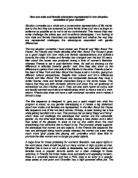
Although physical stimulation is not necessary for an erection it is necessary for semen to be delivered from the penis. Repeated thrust into the vagina of the female leads to the mobilisation of sperm. Muscles encircling the vas deferens contract moving sperm into the urethra. Stimulation eventually leads to the violent contractions of muscles at the base of the penis, resulting in ejaculation (the forceful ejaculation of semen out of the penis). Only a small amount is delivered but it contains several hundred million sperm cells. During ejaculation the sphincter at the base of the bladder is closed, therefore no spermatozoa can enter the bladder or no urine can be delivered. Successful fertilisation requires a high sperm count as the sperm has high odds of making it on the long journey to the egg. A male with a count of less than 20 million per millilitre is considered sterile.
The Female Reproductive System
The main structures of the female reproductive system are the ovaries, the uterus (the womb), the uterine tubes (the fallopian tubes), the vagina and the vulva (external genitalia). Another part that supports the female reproductive system is breast tissue (mammary glands). The female gamete, which contains the genetic information, is called the ovum, egg or egg cell. Ova develop from cells called oocytes located in the outer layer of two compact masses called ovaries, which are contained within the abdomen. Females have at birth all the oocytes they will ever produce.
At ovulation one or a few oocytes initiates development while the others remain in the holding pattern. This long maintenance period is one reason for abnormalities developing with increased frequency in the foetuses of women over the age of 35. Oocytes are exposed to mutation throughout their life and the odds become higher when a woman gets older for this reason.
At birth the female ovaries contain around two million oocytes all of which have begun the first step in maturation and are called primary oocytes. Each primary oocyte is ready to develop further but does not continue to mature until it receives a delivered signal (it is arrested in growth). Very few oocytes do receive this signal of FSH.
Females mature sexually at puberty when FSH starts the3 maturation in a few oocytes, one becomes dominant and the others regress, thus continuing a cycle of 28 days with one oocyte maturing. About 400 of the original 2 million oocytes mature in a woman’s lifetime. When they do they are released from the ovaries and the beating of cilia sweeps the ovum into a fallopian tube, which lead away from the ovary into the uterus. Smooth muscles lining the fallopian tube contract rhythmically moving the ovum down the fallopian tube, the journey is a slow one taking between 3 and 5 days, and if it is not fertilised it loses its capacity to develop after only a few days and once in the uterus it cannot be fertilised at all.
Sperm cells are foreign to the female body and therefore should be recognised and attacked by its immune system like any other foreign body but substances secreted in the semen protect them that inhabit the local immune system. Sperm deposited into the vagina, which is a 7cm muscular tube that leads to the uterus opening, where there is a muscular ring, called the cervix. The uterus is a hollow pear shaped organ about the size of a small fist. The inner wall consists of two layers one that sheds during menstruation and the other inner lining called the endometrium, which regenerates the top layer. Once in the vagina sperm must swim up through the uterus and enter the fallopian tube there is must swim up the fallopian tube and against the current of the tubes contractions, which are carrying the ovum down, and penetrate the membrane of the ovum to fertilise it, during this journey sperm change in ways that help them adhere better to an ovum.
Once the sperm has fertilised the egg high in the fallopian tube it still makes the journey into the uterus and attaches itself to the lining. Once this has taken place many tissues swing into action driven by hormonal signals getting the woman’s body ready, including her womb, to house, nourish and protect the developing foetus. A key structure, which forms from the conceptus, is the placenta, the lifeline between mother and offspring. Through this organ the foetus gains all its needs and disposes of all its waste. Extensive changes occur in almost all of the systems in a woman’s body during pregnancy.
If conception does not occur preparations, in the form of a series of changes each month, are abandoned. The materials that are produced each month are scrapped and a fresh cycle begins, preparing again for conception. This regular sequence is termed the menstrual cycle because of the discharge of fluid (menses) occurring via the vagina at regular intervals. The events of this cycle are divided into four stages: -
1 Menstrual
2 Follicular
3 Ovulatory
4 Luteal
Phase 3, which is mid cycle, is the time that the ovum is released from the ovary (this is what is known as ovulation).