Figure 2 below illustrates the implantation of the blastocyst; - (a) blastocyst adheres to the endometrial lining (b) trophoblastic cells move into endometrium. Disintegration of boundaries between the cells) (c) when implantation finishes the blastocyst is fully buried in the endometrium:
Figure 2 (Human Physiology: from cells to systems- Lauralee Sherwood- 4th edition)
Now the trophoblast cells separate into an inner and outer layer, which is the chorion. The chorion forms finger-like processes called chorionic villi which grow into the endometrium. The arterial and venous vessels inside the endometrium breaks this and let the blood fill the spaces between the chorionic villi. At extremely early stages, nutrient exchange, excretory materials and oxygen between the blastocyst cells and the maternal blood, in uterus pass through the chorionic villi (placenta takes up this function later on.)
After implantation, a cavity, called the amniotic cavity forms in the inner cell mass and causes the part of the inner cell mass near the blastocele to separate as the flat disc of tissue called embryonic disk. The embryonic disk includes two layers of cells: an ectoderm (outer layer) and an endoderm (inner layer.)
At 14 days post fertilization, the embryonic disk becomes slightly elongated oval structure. Some of the ectoderm cells migrate to the middle of the disk, forming a thickened line called the primitive streak. Some of the ectoderm cells migrate through the primitive streak and emerge through the ectoderm and endoderm as a new germ layer, called mesoderm (middle layer). Now the embryo is three layered having ectoderm, endoderm and mesoderm. (Essentials of Anatomy and Physiology – Seeley Stephen Tate – 3rd edition)
Placenta
The placenta is a flat organ that is connected to the uterus on one side and to the embryo, and umbilical cord on the other side.
The needs of the embryo, are met by the placenta, it delivers nutrients and oxygen, absorbs and gets rid of waste products, secretes hormones needed for both maintaining of pregnancy and explusion of the embryo.
Following are the functions of the placenta:
- Delivers nutrients from mother to embryo
- Gas exchange between mother and embryo
- Delivers antibodies from mother to embryo
- Secretions of hormones which include human chorionic gonadotropin(hCG), oestrogen,progesterone, and human chorionic somatomammotropin(hCS)
-
Removal of fetal waste. (Human Physiology Rhoades Pflanzer 4th edition)
Figure 3 below illustrates early embryo and surrounding structures in the placenta:
Figure 3 (Essentials of Anatomy and Physiology – Seeley Stephen Tate – 3rd edition)
Figure 4 below illustrates the relationship between the developing embryo and uterus as pregnancy proceeds:
Figure 4 (Human Physiology: from cells to systems- Lauralee Sherwood- 4th edition)
.
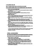
Figure 5 below illustrates the representation of interlocking material and fetal structures which form the placenta:
Figure 5 (Human Physiology: from cells to systems- Lauralee Sherwood -4th edition)
Finger-like processes of chorionic (fetal) tissue form the placental villi. Maternal capillary walls are broken down by the expanding chorion, so the maternal blood moves through the spaces between the placental villi. Maternal blood enters through the maternal arterioles, and then goes through the blood in the intervillus spaces. This is where exchanges are made between the fetal and maternal blood before the fetal blood leaves through the umbilical vein and maternal blood is present through the maternal venules.
The major organ systems start to develop during the embryonic period. This period is also called organogenesis.
I am going to give detail of the face, male / female reproductive tract, heart.
The development of face is by fusion of five masses of tissue. Each mass forms the forehead, nose and middle of the upper jaw and lip. Two of the masses form from the maxillae (upper lip and jaw) and two form the lower lip and jaw. The nose starts as two structures.
When the face reaches maturity and the brain enlarges, the two sides of the nose come to each other in the midline and fuse. The two masses forming the upper jaw expand toward the midline and fuse with part of the nose to form the upper jaw and lip.
The top of the mouth starts to form as ventricle shelves of tissue grow on the inner of the maxillary masses. These shelves swing to a horizontal position and start to fuse with each other at about 56 days of development. (Essentials of Anatomy and Physiology – Seeley Stephen Tate – 3rd edition)
Figure 6 below shows the development of the face:
Figure 6 (Essentials of Anatomy and Physiology – Seeley Stephen Tate – 3rd edition)
The illustration below (figure 7) shows the differentiation of the external genitalia in male and female. At 6 weeks, the genital tubercle, urethral fold, and labioscrotal swelling have differentiated from the genital tubercle (b) At 8 weeks, a distinct phallus is present (c) By week 12, the genitalia have become male or female(e,f) By week 16, the genitalia are formed. (Human Physiology Stuart Ira Fox 5th edition)
Figure 7 (Human Physiology- Stuart Ira Fox - 5th edition)
I have decided to investigate the heart in detail;
The heart is derived from mesoderm; it begins to exist as two simple endothelial tubes which very quickly fuse to form a single chamber or the heart tube that pumps blood by the 22nd day of gestation. Following 2-3 days later the heart tube exhibits four areas, which show the early heart chambers. The four Chambers are:
- Sinus Venosus – this chamber starts to receive all of the venous blood of the embryo. It will become the soft wall part of the right atrium and the coronary sinus. The sinus venosus which gives rise to sinoatrial node which sets the heart rate in the embryonic development.
- Atrium – eventually become muscle ridged parts of the atria.
- Ventricle – is the strongest pumping chamber of the early heart it gives rise to left ventricle.
-
Bulbus Cordis – this chamber gives rise to pulmonary trunk, most of the right ventricle and first bit of the aorta. (Human Anatomy and Physiology Elaine N Marieb 3rd edition)
Figure 8 (Essentials of Anatomy and Physiology – Seeley Stephen Tate – 3rd edition)
The heart undergoes a lot of events and there are major structural changes within next three weeks which results in becoming four chambered organ capable as acting as double pump. Four definitive chambers become part of the heart. Formation of midline septum takes place and pulmonary trunk and ascending aorta form out of bulbus and after this very minor changes occur until birth other than growth.
The interatrial septum of the fetal heart is not complete. The foramen ovale connects the two atria and lets blood enter the right heart to bypass the pulmonary circuit and the collapsed, non functional fetal lungs. Another lung bypass, the ductus arteriosus, is between the pulmonary trunk and the aorta.
Congenital heart defects account for almost for half of the infant deaths arising from congenital anomalies. The most well known abnormalities produce two basic kinds of disorders in the newborn. They either lead to one of the following:
- Lead to mixing of oxygen –poor systemic blood with oxygenated pulmonary blood (inadequately oxygenated blood reaches the body tissues) or
- Involves the narrowed valves that increase the work load of the heart. Examples of the first type of defect are septal defects (shown in the picture) and patent ductus arteriousus, in which the connection of the aorta and pulmonary trunk remains open. A second type of problem could be coarctation of the aorta, whereas tetralogy of fallot. A very serious condition, in which the baby becomes cyanotic within minutes of birth, encompasses both sorts of disorders. Modern surgical techniques can solve most of these heart defects. Most congenital heart problems are traceable to environmental influences, for example maternal infection or drug intake during month 2 when the major events of heart formation take place.
In the absence of congenital heart problems, the heart functions admirably throughout a long lifetime for most people. Homeostatic mechanisms are very effective that it’s difficult for people to notice when the heart is working harder. In people who exercise regularly, the heart adapts to the increased demand by increasing in size and becoming more efficient and powerful pump. The stroke volume increases and resting heart rate declines. Aerobic exercise is also helpful to clear fatty deposits from blood vessel walls throughout the body, so retarding the process of atherosclerosis and coronary heart disease. Barring some chronic illnesses, this useful cardiac response to exercise persists into old age. (Human anatomy and physiology-Elaine N Marieb-3rd edition.)
Figure 9 (Human anatomy and physiology-Elaine N Marieb-3rd edition)
Conclusion
The heart is the most important organ of the body; It is the central organ of the circulatory system acting as a force and suction pump in relation to the blood vessels. Excepting the lungs, it is the only organ in the body through which the blood passes in every cycle.
Congenital heart defects are a problem, but they can be controlled as there is treatment. As the heart is forming defects can occur:
- Ventricular septal defect- there is a fail in the forming of superior part of the interventricular septum.So, the blood from two ventricles mixes. This defect occurs in about 1 in every 500 births.
- transposition of the great vessels-the aorta comes from the right ventricle, and pulmonary trunk from the left. Results when the bulbus cordis doesn't divide in the proper way. The unoxygenated blood passes repeatedly around systemic circuit, where as oxygenated blood recycles around the pulmonary circuit. This defect occurs in about 1 in every 1000 births.
- Coarctation of the aorta- there is narrowing in the part of the aorta, so the work load on the left ventricle increases. This defect occurs about 1 in every 1500 births.
- tetralogy of fallot-has multiple defects; pulmonary trunk narrows and pulmonary valve stenosed; ventricular septal defect; there is opening of aorta from both ventricles; by the overwork wall of right ventricle thickens. This defect occurs about 1 in every 2000 births.
- pulmonary stenosis- there is lessening in the flow of blood to the lungs, because the pulmonary semi- lunar valve is narrowed. This defect occurs about 1 in every 2800 births.
Most of the surgical techniques can correct most of these heart defects.
References:-
-
Human anatomy and physiology-Elaine N Marieb-3rd edition
-
Human Physiology- Stuart Ira Fox -5th edition
-
Human Physiology- Rhoades Pflanzer- 4th edition
-
Essentials of Anatomy and Physiology - Seeley Stephen Tate - 3rd edition
-
Human Physiology: from cells to systems- Lauralee Sherwood -4th edition
- Basic Human Embryology- P.L.Williams /C.P.Vendell Smith
-
Essential Reproduction- Martin H Johnson & Bary- 5th edition
- Human Physiology-Functions of the Human Body- Ross M Durham.