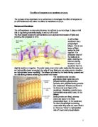
The particles that move through the membrane have kinetic energy. The region with the highest concentration of particles will have the most kinetic energy for that substance. During diffusion the particles move down a concentration gradient until they are spread evenly.
Increasing temperature on the cell membrane means that channel proteins denature and lose their shape. The temperature also increases the kinetic energy of he particles with an increased rate of diffusion. Therefore the rate of facilitated diffusion is also affected and the betalain molecules will leak out and cause the solutions to be darker.
Beetroot contains a pigment called betalain, found in the vacuole which gives the beetroot its characteristic purple colour. Betalain pigments are polar molecules and so are unable to pass through the phospholipid bilayer and can only pass through special channel proteins.
Hypothesis
I predict that as the membrane breaks down, the more pigment will escape and the darker the water solution will become.
Method
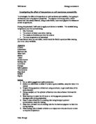
I will need:
- Raw Beetroot
- Size 6 cork borer
- White tile
- Knife
- Ruler
- Water baths
- Beaker
- Boiling tubes
- Racks
- Distilled water
- Pipette
- Stopclock
- Thermometer
- Colorimeter
- Cuvettes
Firstly I will cut sections from a beetroot using a size 6 cork borer. By using a cork borer I will be ensuring the surface area is the same each time, meaning there will be the same amount of membrane structure each time. I will cut 10 sections, avoiding the contact between beetroot and clothing and skin as beetroot dye stains.
Next I will leave the sections in a beaker of distilled water leaving them over night to help the excess dye wash away.
The following day I will place a beetroot section into each of the boiling tubes each containing 5 cm3 of distilled water and place them in water baths at 0°, 10°, 20°, 40°, 60° and 70°. After leaving the boiling tubes in the water baths for 25 minutes the slice will be removed and the boiling tubes shaken.
Afterwards setting up a colorimeter I will set it to % absorbency. I will use a pipette to measure 2cm3 of distilled water into a cuvette and making sure it is placed in with the clear sides correctly I will set it to 0 for clear water.
I will then do the same for each dye solution and test the absorbency at each temperature.
A colorimeter will measure the percentage of light transmission through a solution. This is compared to a cuvette of distilled water, which is the clearest solution.
Variables
The independent variable is the factor that affects the dependent variable, any other variable that may affect the dependent variable must be kept constant.
The independent variable in my experiment will be the temperature and the dependent variable will be the extent of colour change. I will control the size of the beetroot piece and also keep the amount of water they are in constant (5cm3).
Safety
When using the apparatus to cut my beetroot I need to be careful and use a white tile.
While placing my boiling tube into the water baths I will have to be careful not to burn myself and use tongs to take them out. I will also need to take care when using electrical equipment with water nearby. Lab coats may also be worn to prevent dye from splashing and staining the clothes.
Ethics
There are no ethical issues that arise while doing this experiment.
Results
A table to show the percentage light transmission.
My table shows that the average transmission at freezing point is high at 0.08 %, but at 20° the average transmission is only at 0.035% which slowly increases and at 60° is 1.79%. The highest transmission of light was at 70°.
This is probably because the membrane is not designed to exceed its normal temperature. When they do their structures break down which is why my results showed more colour leaked into the water at high temperatures.
Under extreme temperatures the phospholipid bilayer begins to break up due to the kinetic energy gained by the molecules through the heat. This causes gaps in the membrane allowing the fluid to pass through freely without going through facilitated diffusion.
My graph shows a curve. As the temperature increased the channel proteins denatured and lost shape causing betalain molecules to leak out. Also the more I increased the temperature the more kinetic energy the particles had and there was an increase in the rate of diffusion. Putting beet slices in the freezer of course kills them, and afterwards the pigments will leak out.
Evaluation
Overall I think my results were quite accurate and that my experiment went well as they supported my prediction. An increase in temperature denatured channel proteins. To improve the reliability of my results I would repeat my experiment more times to get a more accurate average. I noticed that the most accurate result I got was for the water bath at 70° which I think is because the water bath at 70° had a lid on it, keeping the temperature constant for the whole 25 minutes.
If I was to repeat my experiment I would ensure all the water baths had lids. My points are not all on the line of best fit the most obvious anomaly being 60° , this may be due to experimental errors such as the temperature in the water baths, measuring using beakers and measuring cylinders.
When cutting the beetroot there could have been an uneven distribution of dye and I could have cut through membranes causing further inaccuracy and anomalies. Some piece may have also contained more membrane that others.
There also may have been a lot of inaccuracy when the boiling tube was placed in ice. This is because the ice is constantly melting meaning it is not always at 0°. If I repeated the experiment I would use a freezer so the temperature remained more constant.
Conclusion
I conclude that temperature does affect membrane structure. When the membrane is heated its structure begins to fall apart and the cells are denatured, therefore the colour is darker.