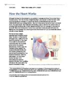
The heart has an own inherent rhythm, that means that the heart muscle, the cardiac muscle, contracts myogenic. This is an independent of external nervous stimulations rhythmic contraction of the cardiac muscle, which is unique for its branched interconnection. The whole contraction of the heart begins at the sinoatrial node, also called the pacemaker of the heart, which is a group of cardiac muscle cells in the wall of the right atrium near the superior vena cava. These cells contract without nevous stimulation. The sinoatrial node spontaneously sends out an electrical signal, which spreads out over the right atrium’s wall into the wall of the left atrium by the Bachmanns bundle. That impulse spread through the atria, causing the atria to contract and to stimulate the atrioventricular node, that is located between the right atrium and the right ventricle. The function of the atrioventricular node is to prevent the ventricles from contracting too early, so that they are full of blood before they contract. This effect increases the efficiency of the heart. After the delay of approximately 0.12 seconds, the impulse is transmitted and via specialized cardiac muscle fibres, the bundle of His, it slowly travels down the septum, where it splits into two branches, the left bundle branch and the right bundle branch. At the bottom of the heart these two bundle branches become thiner and form numerous, small purkinje fibres which spread out the impulse from the bottom to the top and stimulate small groups of cardiac muscle cells. That makes sure that contract from the bottom to the top and not in the middle. This effect is very important because if the heart contracted in the middle, the heart would be much less effective, because not all the blood would be squeezed out of it, but a rest amount would stay in the heart. These electric impulses can be seen in an ECG diagram.
Without further control by nerves, the heart would beat about 100 times per minute. Because of that the heart is connected with the medulla oblongata via two autonomic nerves which send electric impulses to the heart to control the heart rate. The medulla oblongata is a part of the brain and it is located in the brainstem, the lowest part of the brain, which is responsible for most of the essential processes, amongst others for the control of the heart beat. The cardiovascular centre links the vagus nerve and the cardiac nerve with the sinoatrial node. These nerves send either information which cause the heart to contract more frequent or more unfrequent depending on the conditions.
Such conditions are for example a too high level of CO2 in the blood, which can be existant during exercise and which can be decreased by a higher heart rate, change in temperature or in general change in the levels of substances carried in the blood. Such changes are detected by three parts of the body. The first is the carotid artery in the neck, which carries oxygenated blood to the head, the second is the aorta and the third is the medulla oblongata. They send this information to the hypothalamus, a small part of the brain, which is concerned with vital life functions and where the body’s nervous and hormonal systems interact. The hypothalamus then sends signals to either the cardiac or the vagus nerve to speed up or to slow down the heart rate.
When the heart must contract more frequently the cardiac nerve, also called accelerator nerve, is stimulated. The synapses at the end of this nerve secrete noradrenaline, a neurotransmitter, which increases the excitatory postsynaptic potentials, what causes the heart rate to increase. This is the case especially during exercise, when the level of CO2 in the blood increases, so that the rate of CO2 to O2 changes. This control is called negative feedback.
When the heart muscle must contract less frequently, the vagus nerve or decelerator nerve is stimulated and the synapses at the end of this nerve secrete acetylcholine, also a neurotransmitter, which sets up the inhibitory postsynaptic potentials with the result that the heart rate decreases. This is for example the case after doing exercise, when the level of CO2 in blood sinks again or when the body is in rest.
Bibliography:
Michael Kent - Advanced Biology; p.122-125
Andrew Allott, David Mindorff - Biology; p.347-348
Carol Ballard - Body Focus-Heart and blood; p.20
Steve Parker - Body Focus-Brain; p.7, 18, 20-21
Dr. Tony Smith - The human body-an illustrated guide to its structure, function and disorders; p.106-107
Penguin Reference - Dictionary of Biology; p.96, 472, 492