
The biochemical nature oflight detection and emission
The biochemical nature of light detection and emission In this essay I aim to describe the range of biochemical pathways and mechanisms used by living organisms both to detect and to emit light. I will discuss general principles employed, and illustrate the range of different biochemistry involved by the use of many specific examples. Light Detection I will discuss the mechanism and function of light detection by five groups of light detecting molecule. The biggest of these is the rhodopsin group of proteins, I will also look at the role of phytochromes, cryptochromes, flavoproteins and porphirins in light detection. Rhodopsins are found in a diverse array of organisms, all featuring a retinoid prosthetic group linked to a an apo-protein, opsin via a protonated schiff base linkage. Electrons from the schiff base lone pair occupy an extra orbital (the 'n orbital'), therefore electrons can undergo a n-p* transition as well as a p-p* transition. Retinal proteins were first discovered in 1876 by Bell, who observed a reddish pigment that bleaches on exposure to light, which he called visual purple. Most rhodopsins contain retinal as the prosthetic group, but some have one of the other chromophores as shown below. For example freshwater fish have a rhodopsin containing 3,4-didehydroretinal, which has a red shifted UV absorption band. The opsins found in all organisms

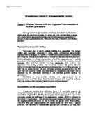
Discuss the stereochemistry of monosaccharides, nucleotides and amino acids
Discuss the stereochemistry of monosaccharides, nucleotides and amino acids. How does the stereochemistry of these building blocks affect the structures of the polymers that they form? Stereoisomers[a] have the same displayed formula but a different arrangement of atoms in space. Enantiomers rotate plane polarised light in opposite ways – each is known as either the D or L form (although this naming originally indicated which way the light was rotated, the fact that this cannot be predicted from atomic structure means that we instead compare the molecule to a similar known one – for example all monosaccharides and amino acids are named based on the structure of glyceraldehyde). The two enantiomers are non-superimposable mirror images of each other - they have a chiral centre (often a carbon atom with four different groups of atoms covalently bonded to it[b]). Monosaccharides[c] Monosaccharides (sugars) are the monomers of polysaccharides (carbohydrates), linked by O-glycosidic bonds which are formed from condensation reactions between the monosaccharides. Monosaccharides have a chiral centre [d]– the carbon one from the end of the chain furthest from the carbonyl group[e]. By convention, the arrangement of atoms in space around this carbon is used to dictate whether the monosaccharide is in its D or L form – although many of the other carbons in the chain are also

An experiment to investigate the change in cell potential with concentration.
8/1/04 Emma Duckworth 7E An experiment to investigate the change in cell potential with concentration. Aim: The purpose of this experiment was to investigate how changing the silver ion concentration in a silver half cell affects the potential of the silver electrode. Apparatus: ==> Chemicals/ substances: * Copper (II) sulphate solution * Copper foil * Silver nitrate solution (make up to 6 different concentrations) * Silver wire * Distilled water * Saturated potassium nitrate solution ==> Additional apparatus: * Safety goggles * 7 beakers * 6 pieces of filter paper * High resistance voltmeter * 2 connecting leads with crocodile clips Diagram: Method: ==> Set up the following cell, using 1 M copper (II) sulphate solution and 0.1 M silver nitrate solution, including a voltmeter and a salt bridge: Cu(s) Cu 2+ ((aq), 1M) Ag+ ((aq), x M) Ag(s) ==> Measure the potential difference of the cell with the voltmeter and note its polarity. Remove the salt bridge as soon as possible. ==> Dilute the 0.1 M silver nitrate solution to 0.01 M silver nitrate solution, renew the salt bridge and then measure the potential difference of the cell with this concentration (0.01 M) of silver nitrate solution in the silver half cell. ==> Repeat this for each of the listed concentrations (0.1 M, 0.01 M, 0.001M, 0.0001 M, 0.003 M, 0.00033 M) of silver nitrate
What are the roles of N- and O-glycans? Use examples to illustrate your answer.
Glycobiology tutorial II: oligosaccharide function Essay 2: What are the roles of N- and O-glycans? Use examples to illustrate your answer. Although the same glycosylation machinery is available to all proteins which enter the secretory pathway in a given cell, most glycoproteins emerge with characteristic glycosylation patterns and heterogeneous populations of glycans at each glycosylation site. What are the roles of these N- an O-linked glycans? Glycosylation and protein folding: The sugars play a role in protein folding and assembly. The proper folding and controlled assembly of many newly synthesized glycoproteins requires them to engage in a series of coordinated interactions with chaperones and enzymes through the attachment of a common oligosacchardide precursor, GlcNAc2Man9Glc3, to N-linked glycosylation sites. This sugar precursor is rapidly processed to GlcNAc2Man9Glc1 which can bind two lectins: the membrane bound calnexin (Clx) and its soluble homolog calreticulin (Clr). Lectins are oligosaccharide binding proteins. The interaction between Clx and/or Clr with nascent monoglycosylated glycoproteins provides access to a folding pathway. In their role as quality factors, Clx and Clr retain unfolded glycoproteins in the ER until they are correctly folded and assembled by chaperones, an event that is signalled by the permanent removal of the terminal glucose
body systems
P3 Body system Cardiovascular system The cardiovascular system is composed of the heart, blood vessels, or vasculature, and the cells and plasma that make up the blood. The blood vessels of the body represent a closed delivery system, which functions to transport blood around the body, circulating substances such as oxygen, carbon dioxide, nutrients, hormones and waste products. There are three main types of blood vessel:- • Veins - the efferent blood vessels that return blood to the heart. • Arteries - the afferent blood vessels that carry blood away from the heart. • Capillaries - narrow, thin-walled blood vessels that form networks within the tissues. Digestive system The digestive system is made up of the digestive tract a series of hollow organs joined in a long, twisting tube from the mouth to the anus and other organs that help the body break down and absorb food. Organs that make up the digestive tract are the mouth, oesophagus, stomach, small intestine, large intestine also called the colon rectum, and anus. Inside these hollow organs is a lining called the mucosa. In the mouth, stomach, and small intestine, the mucosa contains tiny glands that produce juices to help digest food. How digestive system works throughput the body Mouth:-Food enters the body via the mouth where it is chewed. This action helps to break up the food enabling it to be

Describe the structural compartmentation of mammalian cells and the differing functions of these compartments
Describe the structural compartmentation of mammalian cells and the differing functions of these compartments All mammalian cells are eukaryotic cells. They have a true nucleus and they are normally enclosed by a plasma membrane. In a typical eukaryotic cell, one would expect to have, along with a plasma membrane and nucleus, endoplasmic reticulum, Golgi apparatus, mitochondria, cytoskeleton etc.[3] These organelles are all membrane-bound structures, each have a unique role to play in the functioning of the cell. All these organelles are specific proteins and they all interact with each other to support the cell. However cells are different from each other. Cells differ from species to species and they also differ from different organs.[5] Every cell type has its own function and its function determines the quantity of each organelle. For example, we expect to find more mitochondria in a muscle tissue cell than in a skin cell. A muscle tissue cell needs to have extra production of ATP to allow for contraction whereas the skin cell does not need to do so. This assignment will give a brief overview of the structural compartmentation and their function of a typical mammalian cell with special focus on the plasma membrane and mitochondria. Figure 1, a cartoon picture of a typical eukaryotic cell depicting all the compartments[2] Plasma membrane contains the content of the

Skin Cancer
SKIN CANCER Skin cancer is the most common of all the types of cancers (Skin Cancer). It is a disease where malignant, or cancerous cells can be found in some layer of the skin. The epidermis, or the top layer of the kin, has three kinds of cells: basal cells, squamous cells, and the melanocytes (Skin Cancer). The melanocytes produce melanin, which is pigment that gives the skin its colour. Sometimes clusters of melanocytes can form growths called moles. These moles can sometimes become cancerous. Skin cancer tumours are formed when these cells excessively divide. They are known as malignant tumours. The tumours are not skin cancer when they are designated as benign. The cancer cells in the malignant tumours can break away and spread throughout the body. This can occur by way of the blood stream of lymphatic system (Melanoma). Skin cancer is categorized into three types: basal cell carcinoma, squamous cell carcinoma, and malignant melanoma. Basal cell carcinoma is, by far, the most common form of skin cancer (Skin Cancer). Luckily, it will never spread throughout the body. Basal cell skin cancer usually forms due to years and years of skin damage. Therefore, it is most often found on the sun-exposed areas of older adults. Such areas include the nose, face, back, and neck. Basal cell carcinoma is definitely curable with treatment. The second most common
The Use of Controls and Indicators to Find What Macromolecules are in an Unknown
The Use of Controls and Indicators to Find What Macromolecules are in an Unknown Name: Erin Teaching Assistants: Laura and Polina, Section # 013 Course # 4677 Day: Monday, Time: 7:00PM - 9:50PM, Room #: B2 151 Lab Performed: Monday September 15th, 2008 Introduction The objective of this lab was to discover what macro molecules were in the unknown using different substances as experimental controls. Experimental controls are samples used in a lab that have a known outcome. This outcome can show what an unknown is comprised of. A positive control shows what the outcome of the experiment should look like if the experiment were to work. A negative control shows what would happen to the substance if the experiment were to go wrong. When compared to the control, what type of macromolecules in the unknown can be discovered (Enlexica, Inc, 2008). Indicators are usually used to expose a certain substance within a substance and normally shown through a colour change. These indicators were used to create the controls and used to identify certain macro molecules in different substances. The three different indicators that were used in this lab were Lugol's iodine solution, Benedict's solution and Biuret solution (Buckley Jr., 2003). Lugol's solution, otherwise known as iodine solution is an indicator used to find starch in a substance. This solution is made of 5% iodine, 10%

Osmosis - diffusion - cemi permeable membrane
All particles are in constant motion. In solid particles (ions, molecules, atoms etc) vibrate. In liquids they move more easily, while in gases they move randomly. Particles have a natural tendency to move from areas of their higher concentrations to areas of their lower concentrations. This is called 'DIFFUSION'; it is a passive process and does not need the use of energy. How quickly it happens depend on the difference between concentrations. OSMOSIS is a special case of diffusion where only water molecules are involved. It is the movement of water molecules from a region of higher water potential to a region of lower water potential across a PARTIALLY PERMEABLE MEMBRANE (1). Partially permeable membranes allow only small particles to flow through them. Living cells' membranes possess the same characteristics to control exchange of materials for their survival. The task is to find out the water potential of potato. This can be obtained by putting potato pieces in different water concentrations. The equipments that I will use are: * 50 cm3 beakers (5) * 2 potatoes * 25 cm3 measuring cylinder (2) * Cork borer (1) * A knife * Electric balance * 2 molar sucrose solution (1): CAMBRIDGE ADVANCED SCIENCES. BIOLOGY 1 PUBLISHED BY THE PRESS SYNDICATE OF THE UNIVERSITY OF CAMBRIDGE. Page 55-56. The following diagram shows the motion of water particles from

Are CD46, CD55 and CD59 good targets for the treatment of malignant disease?
BM301 Fundamental Immunology | Essay Fundamental Immunology: Are CD46 - CD55 - CD59 good targets for the treatment of malignant disease? Are CD46 - CD55 - CD59 good targets for the treatment of malignant disease? Clusters of differentiation which is often abbreviated to CDxx (xx representing a number specific to each CD molecule), is a naming system developed in 1982 to recognise surface molecules present on white blood cells that were recognised by monoclonal antibodies (Fiebig et al., 1984). They can perform a number of functions such as acting as receptors or ligands and are usually involved in cell signalling, causing a cascade in certain immune responses. There are now over 250 proteins that are classified as CD molecules (Zola et al., 2005). This essay will look at three clusters of differentiation - CD46, CD55 and CD59 - known membrane proteins that protect against native complement damage (Xu et al, 2008) and conclude whether or not they are good targets in the treatment of malignant disease. CD46 (Membrane Cofactor Protein) is a type I membrane protein which acts as a complement receptor. It is a cofactor for the proteolysis of C3, so cells displaying CD46 will be protected from attack by the native complement system (Assem et al., 2005). CD55, which is also known as Decay Accelerating Factor is also a membrane protein with a similar function to CD46. CD55 causes