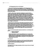
Statisticians knew that some blood types were more common, or more common in certain ethnic groups than others, and tried to bring mathematics to blood typing (see charts one, two and three, page 16 and 17) ( and http://www.clinical-blood-testing.com/blood_typing.htm ). Blood typing remained circumstantial evidence, but it wasn’t going to convict anyone on its own. Unlike fingerprints, which were unique to an individual, blood types could be shared by millions of people. Other biological clues like the Barr bodies, a piece of chromosomal material in the nuclear membrane of female cells which is the remnant of one of the two female chromosomes, found in the cells of females might help narrow the pool of suspects, but even half a million people is still a pretty big pool.
For that kind of individualization, law enforcement had to wait until DNA analysis, the study of deoxyribonucleic acid, advanced. DNA, the physical material that we inherit from our parents when that one sperm finds that one egg, is absolutely individual-with one exception. Identical twins form from one fertilized egg, so their DNA is identical (Microsoft Encarta Encyclopedia, 2003).
While fingerprinting is still the only surefire way to separate identical twins, DNA testing provides its own advantages.
Every cell in an individual’s body contains identical DNA. Fingerprints come only from fingers, but DNA can be found in blood, in urine, in feces, in saliva, in some hair, in the shed skin cells found in a facecloth or toothbrush, even the sweatband of a hat (). A suspect doesn’t have to bleed at the scene to leave DNA. Semen at rape scenes, saliva on the envelope of a ransom note, skin cells scraped onto a rope while tying up a victim, all provides the opportunity for collection and analysis.
DNA can survive much longer than a fingerprint. While some few prints have been collected years after being made, DNA analysis has been done on Egyptian mummies. DNA can provide closure to a victim’s family years latter by making it possible to identify bodies that might have been buried a decade ago as John or Jane Doe.
DNA can indicate familial relationships. Though it’s been theorized that prints in family members might have similarities, it’s unproven. But because DNA is inherited from two parents, a significant number of matches in a sample can point to a “first-degree” relative-a mother, father, or sibling. In cases where groups are involved in crimes, this is important evidence.
DNA evidence doesn’t combine. Blood evidence at a scene frequently comes from more than one individual; either the attacker and the victim or a number of victims. If one hypothetical victim is type A and another is type B, a combined sample of their blood might suggest an AB individual. With DNA, the traits of both victims would be found in the sample. But with samples from the victims themselves available for comparison, it would be possible to prove that it’s a combined sample possible from only these two individuals (Evett, I.W. & B.S. Weir., 1998). It makes determining who was at a specific point in the scene possible.
With so much at stake, collecting DNA evidence at the scene, as well as from both victims and suspects, is imperative. But it’s not always easy. Collecting and preserving quality DNA samples requires that investigators first determine where DNA samples might be found, and then to ensure that it is collected without any contamination occurring to what may well be a tiny sample.
Before it can be collected, it must be found. And if one of DNA’s advantages to the investigator is that small samples are still useful, then one of its disadvantages is how hard to spot such tiny deposits can be. Two of the aids investigators may use are chemicals like luminal and alternative light sources like UV. Many biological samples fluoresce naturally in certain wavelengths of light. Semen, blood, and amniotic fluid are amenable to this method. If it’s not possible to use an alternative light source, a spray of luminal (which reacts with the iron in blood and any other blood containing biological specimens) proves valuable.
In the United States, the National Institute of Justice has a checklist of items investigators should consider in determining where DNA evidence might be found and suggests the collection of these:
- Fingernails or fingernail clippings.
- Tissues, paper towels, napkins, cotton swabs (bag everything in a bathroom wastebasket).
- Toothpicks, cigarette butts, straws, anything else that might have been in contact with the mouth like cellular phones.
- Blankets, pillows, sheets, mattresses, dirty laundry.
- Head gear of any type.
- Eyeglasses, contact lenses.
- Used stamps, envelopes.
- Tapes, ropes, cords, anything else used as ligatures.
- Used condoms.
- Bullets that have passed through bodies.
().
Clearly many of these sources will be submitted for more than DNA examination, so it’s essential that prioritization and joint handling procedures be agreed upon by all investigation members to avoid loss of evidence.
Since DNA is found everywhere, extraordinary efforts must be made to isolate samples from each other and to prevent investigators from bringing in inadvertent samples. To avoid contamination of evidence that may contain DNA, always take the following precautions:
- Wear gloves. Change them often.
- Use disposable instruments or clean them thoroughly before and after handling each sample.
- Avoid touching the area where you believe DNA may exist.
- Avoid talking, sneezing, and coughing over evidence.
- Avoid touching your face, nose, and mouth when collecting and packaging evidence.
- Air-dry evidence thoroughly before packaging.
- Put evidence into new paper bags or envelopes, not into plastic bags. Do not use staples.
().
All evidence collection should be undertaken in as timely a manner as possible, especially if biological evidence is involved. Humidity, direct sunlight, rain, and the growth of bacteria can all degrade samples beyond recovery. And removing the evidence to the inside of an evidence van sitting in bright sunlight doesn’t solve the problem, it just bakes the evidence. So air conditioned or air controlled containers are essential equipment.
The flip side of the contamination issue is that DNA sampling, which brings investigators into proximity with any number of bodily fluids, requires special precautions to ensure the investigator isn’t contaminated by the samples. Biohazards like hepatitis and HIV are real dangers to investigators and laboratory personnel who handle bio-wastes and bio-fluids, so all samples must be treated as infectious until proven otherwise.
DNA permeates every cell of the human body, but investigators are limited to direct sampling of body cells or body fluids. Blood recovered from flea, cockroach and mosquito guts can and has been successfully submitted for ABO and DNA typing. Human blood in cockroach feces turns up under luminal testing and has actually confused the blood splatter analysis when fecal matter around a room has been mistaken for dried blood droplets ().
With a good uncontaminated sample that hasn’t been baked or grown moldy, lab personnel can decide on a typing technique. The earliest DNA typing was the RFLP, restriction fragment length polymorphisms, method. In this technique, DNA extracted from the sample is cut into fragments by chemical “scissors,” which separate the long DNA strands at specific spots between any two of the four proteins that make up DNA. These four proteins are adenine (A), thymine (T), cytosine (C), and guanine (G). Because everyone’s DNA is different, the length between one persons A’s and T’s or C’s and G’s is different from the distances between another persons.
Suppose we have two suspects and a pair of chemical scissors that will snip every C to G bond in a string of DNA proteins.
In suspect one, CGATTAGCGAGCT becomes C GATAG C GAG CT.
In suspect two, TTCGTATATATACG becomes TTC GTATATATAC G.
There are some vary short pieces, some medium length pieces, and some very long pieces.
Next, a suspension gel that can carry an electrical charge is set up. This part of the process is called gel electrophoresis. DNA from each person, a control sample, and the sample from the unknown are set into different lanes of the gel so the material doesn’t get mixed. Then current is run through the gel. When that happens, the fragments begin to line up. Lighter pieces (the shorter ones) travel much farther than the heavier (longer) pieces and, when the process is complete and every thing is at rest, the fragments will have formed into distinct bands based on their weight (see chart 4, page 17 ). These bands become visible when they’re tagged and x-rayed. It’s usually these films that are seen in court.
If two samples come from the same person, they’ll break along the same lines and come to rest in the same places. That makes a match. If they aren’t in the same place, there’s no match. If there are considerable similarities but no match, it’s possible the samples are from related individuals.
The RFLP technique is a detailed, accurate, and precise way of identifying individual people. On the downside, RFLP is slow. It can take anywhere from three weeks to three months to get results. The lab work alone takes nearly a month and, because it ties up lab facilities, there’s frequently a backlog of work. Not helpful if you’re a law enforcement officer who needs a lead and certainly not helpful if you’re the accused waiting for these results to clear you. On top of that, RFLP requires a lot of fresh sample. Large blood splatters, hair samples of up to twenty-five hairs, or large quantities of saliva are needed for each test. A semen sample the size of a large thumbnail may not contain enough DNA for testing. If you only have one sample, this method might take up all the available raw material.
PCR, polymerase chain reaction, methods improved on the RFLP technique by replicating the DNA present in a tiny sample until there was enough of it to type, making even minuscule samples significant. It works on the principle that DNA replicates itself naturally each time a cell divides. PCR creates the same situation chemically by “unzipping” the DNA molecule into two halves. Because the exposed molecule ends on each can only attach to their opposite chemical numbers (supplied in raw form by the technician) the created half is identical to the original. When both halves have grown new halves, the amount of identical DNA has been effectively doubled. And the process can be repeated by unzipping the new bits and allowing the chemical reactions that create new halves to recur as often as if needed to obtain a large enough sample. From that sample, it’s possible to compare evidence based on the locations of specific markers on just a single stretch of DNA. The results from PCR testing of this type are seen as a series of dots. If the dots are darker than the control dot, or C dot, it’s a match; if not, it’s not.
The advantages of PCR are many. To begin with, it’s fast. Even the longest testing period should take less than a week, and it’s frequently back faster than that. Some preliminary results can come back in forty-eight hours. The whole process of duplication occurs in a single vial that contains the sample to be copied, lots of raw material for the natural duplication process, and some primer (the chemical that starts the duplication reaction). The vial is first heated to about 160° F for thirty seconds. This allows the two halves to unzip. When it is cooled to about 100° F, the primer kicks in and, in less than half a minute, the process of duplication begins. The vial is reheated to about 140° F and duplication continues until both halves of the strand have been copied. The whole process takes about two minutes and it’s repeatable. In less than four hours, it’s possible to multiply a sample by 5,000 percent (see chart five, page 18).
Also, PCR testing is much less expensive than RFLP methods, making it possible for more case evidence to get into your system.
PCR can also use degraded evidence; in other words, older samples recovered at a secondary scene located some time later, even up to decades after the primary scene has been worked and, can still produce significant results. Tiny samples, a single hair, blood or semen stains smaller than the ball of a ballpoint pen, are sufficient.
Unfortunately, PCR samples are at higher risk for contamination than are RFLP samples. The copying process can’t distinguish between DNA left by a suspect and DNA from a criminalist who breathed a little too heavily; consequently, all DNA present is copied. From a purely statistical perspective, because of the narrower region of study, the results may not be as detailed as RFLP. One in millions, or even billions, are terms associated with RFLP; with PCR, the numbers are more like one in ten thousand or one hundred thousand- impressive, but not of the same order.
DNA analysis is a quickly changing field. Just when PCR, its values and differences from RFLP, was becoming generally understood, new tests, the STR (short tandem repeat) process in particular, began yielding significant results. STR’s are newcomers in court, but have been used extensively in non forensic identifications. Victims of Flight 800’s crash off Long Island were identified with this method as were those who died in Branch Davidian fire outside Waco, Texas. It seems likely that the victims of the September 11 assaults may be identified by the same methodology. As might be inferred by these incidents, STR techniques can function with severely degraded DNA.
The small amount of DNA available is multiplied by using the PCR technique, but the raw material used in the duplication process has already tagged with fluorescent dyes. When the gel electrophoresis process is complete, the bands can be read with lasers instead of X-rays, and the analysis of the locations can be read by a computer. Multiple samples can be run at once, and the results are a considerably more readable graph, the electropherogram, instead of X-ray films (see charts six and seven, pages 19 and 20).
DNA analysis, frequently call DNA fingerprinting, does share one other characteristic with the more established science of fingerprinting- its results can be coded for storage and retrieval in a database. STR’s are easiest of all. The genetic equivalent of AFIS (Automated Fingerprint Identification System) is CODIS (Combined DNA Index System).
CODIS is used in the national, state, and local index system networks to link typing results from unsolved crimes with cases in multiple jurisdictions or persons convicted of offenses specified in the data banking laws passed by the jurisdictions. By alerting investigators to similarities among unsolved crimes, CODIS can aide in apprehending perpetrators who commit a series of crimes.
Bibliography
Alonso, K. & C. (1997) Forensic Pathology. Atlanta, Georgia: Allegro Press.
Blackett Family DNA Activity. Retrieved September 22, 2003, from http://www.biology.arizona.edu/human_bio/activities/blackett/anatomy.html.
Blood Typing. Retrieved September 21, 2003, from http://www.clinical-blood-testing.com/blood_typing.html.
Blood Typing Facts and Statistics. Retrieved September 21, 2003, from http://www.craigmedical.com/blood_typing_facts.html
Dix, J. (1998) Guide to Forensic Pathology. Boca Raton, Florida: CRC Press.
DNA Fingerprinting. (2003). Microsoft Encarta Encyclopedia. Microsoft Corporation.
Evett, I.W. & B.S. Weir. (1998) Interpreting DNA Evidence: Statistical Genetics for Forensic Scientists. Sutherland, Mass.: Sinauer Associates.
Polymerase Chain Reaction. Retrieved September 22, 2003, from .
Rh Factor. (2003). Microsoft Encarta Encyclopedia. Microsoft Corporation.
Welcome to Earl’s Forensic Page. Retrieved September 22, 2003, from http://members.aol.com/EarlNMeyer/DNA.html.
. Retrieved September 21, 2003 from .
What is Forensic Entomology? Retrieved September 21, 2003 from .
Chart One
Blood Type frequency in percentage of total population:
Chart Two
The overall statistical distribution of blood type plus Rh factor in the total population is as follows:
Note: Percentage distribution may be different within specific racial and ethnic subgroup. (See chart three) (Charts one and two are from http://www.craigmedical.com/blood_typing_facts.html)
Chart Three
Since blood type is an inherited characteristic, different ethnic groups have different proportions of the basic blood types. The following table shows these percentages for different ethnic groups in the United States. Most people (about 85%) have Rh-positive blood.
()
Chart Four
(http://www.biology.arizona.edu/human_bio/activities/blackett/anatomy.html)
Chart Five
(http://www.accessexcellence.org/AB/GG/polymerase.html)
Chart Six
(http://members.aol.com/EarlNMeyer/DNA.html)
Chart Seven
(http://members.aol.com/EarlNMeyer/DNA.html)