One important limitation in this study is that the group’s data must be interpreted in a manner such that the development of cardiac hypertrophy, following acute pressure overload, is different from cardiac hypertrophy, which occurrence is secondary to heart diseases in the clinical setting. Therefore, the experimental group’s mouse model of hypertrophy could be examined as their main advantage and limitation. They decided to choose TAC rather than ascending AC because this allowed a low resistance outlet.35,37,38 However, according to histological examinations, the hypertrophied ventricles only contained small localized foci of cardiac damage, which account for less than 5% of the ventricle.35,39 One of the main advantages of this study is the examination of whether pressure overload involving a specific interaction with βAR signaling pathway causes increased GRK2 activity. A genetic model of cellular hypertrophy was used in order to establish that increased GRK2 with pressure overload hypertrophy was a cellular response of increased protein synthesis, which further induces cardiac hypertrophy.35
The overall implication asserted by Choi et al. is that GRK2 is responsible for altered βAR signaling associated with HF, i.e. cardiac hypertrophy. The study explains how increased GRK2 accounts for cardiac hypertrophy via the uncoupling of βARs. Furthermore, βAR desensitization does occur with the development of pressure overload cardiac hypertrophy.35
The possibility of increased GRK2 mRNA was further investigated in a study by Harris et al.17 This group’s purpose was to establish the possibility of increased GRK2 in cardiomyopathic hamster tissue and monitor its expression during development of HF. Therefore, Harris et al. tested the hypothesis that GRK2 expression increases in the CHF 147 LV during progression of HF. In this study, Harris et al analyzed GRK2 levels in the cardiomyopathic Syrian hamster CHF 147’s cardiac tissue and compared the levels to the non-cardiomyopathic control hamster (CHF 148). The methods utilized in this experimental study were: (1) ion exchange chromatography to obtain purified GRK2. (2) Phosphorylation of rhodopsin to assay GRK2 activity, (3) Anti-GRK2 polyclonal antibody to detect GRK2, (4) Mass spectrometry to analyze GRK2 sample, (5) Densitometric scanning of the autoradiographs to quantify results from the cardiomyopathic samples, and (6) Protein extracts from hamster lymphocytes were used to recognize hamster GRK2; which was further analyzed by western blotting.17
Harris et al. reported: (1) an increased expression of GRK2 rat brain compared to the hamster lymphocyte sample, (2) Greater expression levels of GRK2 in cardiomyopathic samples compared to the controls of 100,180, and 350-day-old samples but not for those at 30 days, (3) significant increase in GRK2 expression by 2.0-, 2.56-, and 4.48/5.01-fold in cardiomyopathic tissue as compared to the normal tissue at 100,180, and 350 days, (4) a 2.2-fold increase in GRK2 band intensity in cardiomyopathic tissue and (5) a 2.56 fold increase in cytosolic fraction when GRK2 expression was analyzed in the membrane fraction from the 180-day LV samples.17
The Syrian cardiomyopathic hamster CHF 147 is an animal model of dilated HF.17,40 This animal model further supports the results because in humans, this model is analogous to severe stages of HF when βARs are downregulated.17,41 So this served as an advantage for the group. However, there are many limitations that need to be taken into consideration. The most important of these is the fact that GRK2 activity was not detectable with rhodopsin because the levels were too low in the cardiac tissue. The immunoreactive bands did not clearly visualize GRK2 in a 300μg heart-derived protein on a gel compared to 10μg brain or lymphocyte protein extracts. The loss of GRK activity upon freeze-thawing the tissue can be considered another limitation. This may lead to decreased levels of GRK2 expression compared to the mass of the protein, which may further affect the results the group has reported.17 Furthermore, Ping et al reported constant GRK2 mRNA and protein level following pacing-induced CHF in pigs; however GRK5 mRNA and protein had increased.17,42 In this instance, these results do contradict the results reported by Harris et al. Therefore some variability may exist with respect to whether GRK2 or GRK5 expression increased depending on the different animal models used for HF. This could further imply that different species undergo different methods of progression towards HF.17
However, in humans, GRK2 in HF has been shown; therefore the study performed by Harris et al. may be beneficial in understanding the development of HF via increased GRK2 expression. Furthermore, these animal models should be useful in testing the effect of GRK2 inhibitors in the development of HF. Thus, the Harris et al. data does support a key role for increased GRK2 expression in the development of HF in CHF 147 cardiomyopathic hamsters.17
It is well known that the characteristic changes in myocardial expression of GRK2 are associated with the onset of HF. However GRK2 expression during cardiac hypertrophy without HF had not been explored until 2002, by Theilade et al. This group’s purpose was to indicate differential GRK2 expression during cardiac hypertrophy with and without HF in response to myocardial infarction (MI) in rats. Furthermore, this group tested the hypothesis that GRK2 expression is differentially regulated in these two groups of hypertrophic hearts.43
The experimental study was carried out using various techniques discussed above, such as western blotting, anti-GRK2 antibody, cardiac catheterization. Moreover, the rats subject to experimental studies had an occluded left anterior descending coronary artery. This ligature was not closed in the sham-operated animals. Arterial pressure, LVEDP (left ventricular end-diastolic pressure) and maximal rates of isovolumetric pressure development (dP/dtmax) and decay (dP/dtmin) were measured through cannulation of the right carotid artery. Infarction sizes within the ventricles LVVEDP was used as a control. Casein assay was used to detect GRK2 activity. Full speed centrifugation was used to remove insoluble material from the myocardial tissue placed in a buffer solution.43
GRK2 expression was tested in hypertrophic hearts with and without HF in response to MI. Later on, the post-MI rats were divided into two groups according to the absence or presence of pulmonary edema. The following results came from this study: (1) a significant increase was observed in the RV mass index at three and nine weeks after MI; the group with hypertrophy and HF had pulmonary edema, (2) animals with HF exhibited an increased LVEDP; animals without HF had reduced dP/dtmax and dP/dtmin, in contrast to those with HF, (3) western blot indicated that GRK2 expression was inhibited in animals without HF while it was enhanced in those with HF, (4) GRK2 activity increased by 1.32 in the group with HF, compared to the sham-operated group; GRK2 activity decreased by 0.58 in the group without HF. Furthermore, the differential GRK2 regulation was still present nine weeks post-MI. GRK2 activity in animals with HF was increased by 1.21, while it was suppressed to 0.62 fold of control values within animals with myocardial hypertrophy.43
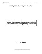
Figure 5: (a) Overexpression of the GRK2 gene was achieved by incubation for 48 h after infection with an adenovirus containing the GRK2 gene, and cardiomyocyte lysates were analyzed by western blotting. (b) Homogenates from the myocardium of rats were analyzed by western blotting three weeks after experimental infarction. Animals with infarctions were divided into two groups, animals with hypertrophy respectively animals with hypertrophy and pulmonary edema (heart failure), and compared to sham-operated animals. (c) Western blots are presented as a bar histogram. Error bars indicate SEM. P-value was derived from an ANOVA.43
Figure 6: (a) GRK2 activity in the myocardium was analyzed 3 weeks after experimental myocardial infarction. Animals were divided into two groups depending on thepresence of pulmonary edema as a sign of heart failure, and the groups were compared to sham-operated animals. (B) Graphical representation. Error bars indicate SEM. P-value < 0.01 in a Kruskal-Wallis test. 43
Figure 7: (a) GRK2 activity in the myocardium was analyzed 9 weeks after experimental myocardial infarction. Animals were divided into two groups depending on the presence of pulmonary edema as a sign of heart failure, and the groups were compared to sham operated animals. (B) Graphic representation of GRK2 activity. Error bars indicate SEM. P-value < 0.05 in Kruskal-Wallis a test
A thorough analysis was performed by the group to prove their hypothesis correct. They were successful in demonstrating GRK2 as an important regulator of cardiac function. However many limitations can be identified as well. As it was reported in the study by Harris et al, there may be different GRK2 regulation between different species of animals. Therefore, the consequence of GRK2 levels associated with the possible risk of HF needs to be further researched. In many cases, GRK2 upregulation may be a precipitating factor. There are also other factors that regulate GRK2 and its progression to HF as well. Therefore, the signaling systems in the hypertrophic heart with and without HF post-MI’s signaling pathways should be taken into consideration and compared.43
The group’s findings have demonstrated a strong correlation between HF and GRK2 upregulation. Furthermore, the GRK2 levels were dependent on the presence of HF after MI. Reduced GRK2 levels was observed in animals with cardiac hypertrophy without HF, while animals with HF had elevated GRK2 expression and activity. Therefore, their data suggested differential regulation of GRK2 occurs in hypertrophic hearts with no HF and with HF. Animals without HF had inhibited expression and activity of GRK2 while those with HF had elevated levels.43
GRK2 interacts with several intracellular molecules, which contribute to its regulation by affecting its subcellular localization.44 The consequences of these interactions are not fully understood but they do carry some significant importance with respect to its signaling and regulation.44 Moreover, GRK2 activity has been seen to inhibit and activate many signaling systems in cardiomyocytes.44,45 This has raised many questions regarding its physiological and pathological role in the heart. The enhanced GRK2 activity and expression observed in various animal models of HF suggests that its activity may explain the concurrent downregulation of βAR.44.46 Myocardial GRK2 expression in mice can be changed with transgenic technology.44 GRK2 overexpression results in reduced cardiac contractility while GRK2 KO proves lethal in utero.44,36 However, GRK2 inhibition via overexpression of GRK2-ct has shown such beneficial effects as reduction in hypertrophy and increased survival.44 Furthermore, GRK2 inhibition has been shown to increase βARs on cells surface.44,8 Therefore, GRK2 inhibition may be an interesting therapeutic potential for increased receptor signaling.44
GRK2-ct in infracted rabbit hearts has revealed improved cardiac function.47 However, the possibility of improved cardiac function induced by MI was not studied until 2004. Suzuki et al. studied WT and TG mice with an overexpression of GRK2-ct following MI. The purpose of this study was to determine whether inhibition of GRK2 would improve cardiac function and further survival in mice with HF. This group hypothesized that GRK2 inhibition would increase survival and improve cardiac function in these mice according to the results of previous experimentation studies of gene therapy using GRK2-ct.47
The methods employed by this group were transthoracic echocardiography to measure LVEDD, LVESD, AWth, PWth; 125I-cyanopindolol in binding buffer with membrane fractions determined myocardial βAR density; myocardial GRK2 protein levels were determined using polyclonal antibodies and chemiluminescence conjugated with anti-rabbit IgG. The TG mice were a cross of heterozygote GRK2-ct and WT. In this study, MI was produced by permanent ligation of the left coronary artery. Sham operated mice were subject to similar surgeries except for an occlusion of the coronary artery. The group chose to exclude mice with MI, which died within 24 hours or a week for survival analysis.47
The following results emerged from this study: (1) At 26 weeks, survival rate of WT mice with MI was 25% while GRK2-ct TG mice was 92%; the survival rate was not due to infarct size since no differences were reported; (2) at 8 weeks after MI, echocardiography revealed an increased LVEDD and LVESD in WT and TG, compared to the sham operated mice; (3) cardiac hypertrophy, LV dilation and cardiac dysfunction was observed in mice with MI, while GRK2-ct TG mice exhibited a smaller LVESD; (4) reversed βAR downregulation due to an overexpression of GRK2-ct, and (5) WT mice with MI had higher cytosolic myocardial GRK2 levels compared to sham-operated WT mice, and (6) GRK2 levels were similar for GRK2-ct TG mice with MI had similar levels to the sham-operated WT.47
The development of HF induced by MI is indicated by βAR signaling abnormalities such as βAR downregulation and increased myocardial GRK2 protein levels. Furthermore, the fact that animals18,35,47,48,49, with congestive HF and humans with HF32,45 exhibit similar outcomes serves as a main advantage for the group and for the future direction of this research. However, the main limitation associated with this study is that the mice were only monitored for survival for the first 26 weeks after MI, since the long-term mortality rate of the mouse model was not known. Moreover, this study establishes improvement in survival of HF induced MI mice through expression of GRK2-ct peptide. Cardiac dysfunction and βAR signaling abnormalities after MI are inhibited by GRK2-ct.47
One important limitation that needs to be taken into consideration is the fact that βAR signals to various other effectors through diverse intracellular proteins. The fact that chronic βAR downregulation and desensitization can attenuate other intracellular pathways (i.e. cell growth) may indicate that βAR downregulation-dependent cell growth signaling pathways are not involved in pathological progression to HF. Furthermore, cardiac hypertrophy after MI was not attenuated by myocardial GRK2-ct expression in GRK2-ct TG mice with pressure overload cardiac hypertrophy.47
Moreover, the study reveals increased survival, improved βAR signaling and delayed progression of cardiac dysfunction in the mouse model of HF induced by MI. The results support the idea that increased GRK2 activity and βAR desensitization may begin as an adaptive mechanism and later on proves detrimental to HF. In addition, myocardial GRK2 inhibition restores βAR signaling. Therefore, GRK2 inhibition may represent a therapeutic approach for HF.47
It can be postulated that a chronic increase in CA in the heart causes βAR dysregulation, which further results in many molecular abnormalities including upregulation of GRK2.20,8 α2ARs mediate almost all presynaptic inhibitory autoreceptor function and normal SNS regulation and function.20 Furthermore, autocrine feedback inhibition of CA secretion from the adrenal gland is exclusively mediated by α2ARs. 20,30,31 A study involving genetic KO of individual α2ARs in mice may shed some light on the role of sympatho-inhibitory α2ARs in the regulation of cardiac function. Individual and double KO mice of α2AAR- and α2CAR was characterized by enhanced SNS activity and circulating CA levels, worsened heart function after surgical pressure compared to the stressed control mice.20,25,26,27 Therefore, this indicates that regulation of SNS activity and CA outflow may be mediated by α2ARs, including those in the adrenal gland. However, the relation of HF and the importance of adrenal α2ARs for SNS function had not been tested until 2007.20
In previous experiments, no researchers have emphasized adrenal α2ARs and their importance in SNS function, their role in adrenal physiology and CA secretion in HF. A study conducted by Lymperopoulos et al. in 2007 investigated the overexpression of GRK2 and the function of the adrenal gland in failing hearts. This group used two different HF models to test the adrenal adrenergic receptor system and its regulation. The objective of this study was to reveal that dysfunctional adrenal α2AR signaling and SNS hyperactivity and increased CA secretion in HF is due to GRK2 upregulation. Furthermore, the group hypothesized that adrenal GRK2 inhibition may be a potential sympatholytic strategy for the treatment of HF.20
The two different HF models employed by the group were TG mice with cardiac overexpression of calsequestrin (CSQ-TG mice) and rats with chronic HF following MI. CSQ is a sarcoplasmic reticulum calcium-binding protein. Their cardiac function was evaluated based on the results of echocardiography and LV catheterization. The following other techniques were utilized to derive results: (1) Isolation and culturing of adrenal chromaffin cells; immunoblotting and immunofluorescence to purify the cultured adrenal chromaffin cells, (2) in vivo adrenal gland delivery; (3) ELISA to measure plasma and in vitro CA secretions; (4) RNA isolation, reverse transcription and quantitative real-time PCR to measure mRNA levels of α2ARs in the adrenal gland; (5) western blotting, and (6) saturation ligand-binding using the expression for tyrosine hydroxylase.20,6,51 Tyrosine hydroxylase is an enzyme that catalyzes the rate-limiting step of CA synthesis in chromaffin cells.20
The functional status of the adrenal medulla was examined first, since the adrenal medulla is the key SNS component for overall CA outflow. CSQ-TG and HF rats had considerable adrenal hypertrophy and elevated expression of tyrosine hydroxylase (mRNA and protein) in comparison to the control group (NLC). CA secretion from the adrenal medulla is solely inhibited by α2ARs. No differences in mRNA were reported for the control and CSQ-TG and the sham-operated mice and the experimental group. A ∼40% decrease in α2AR density in CSQ-TG compared to NLC group was reported. This established the occurrence of substantial adrenal α2AR downregulation at the post-transcriptional level in HF. These results were independent of the species and α2AR studied. This can be noted as an advantage of the study since many others in the past have reported variability amongst species.20
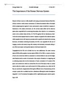
To ensure the action of GRK2 is associated with the downregulation of α2AR, the expressions of endogenous GRK2, 3, and 5 were studied at the mRNA level.20,0 An increase in adrenal GRK2 occurred in both models of HF compared to the control, while GRK3 and GRK5 levels remained fairly stable. This aspect of the study further helped the group establish their hypothesis that it is indeed the actions of GRK2 which lead to α2AR downregulation.
Figure 8: Adrenal GRK2 upregulation in heart failure. (c) Western blotting for GRK2 in adrenals of these mice (left) and rats (right).20
GRK2 protein is present in adrenal medulla chromaffin cells. Its presence has been confirmed by immunoblotting and immunofluorescence. Furthermore, co-immunofluorescence established GRK2 presence through the process of tyrosine hydroxylase co-localization. Both models of HF exhibited an elevated level of GRK2 compared to controls. These observations indicate upregulation of GRK2 in HF, which may further trigger downregulation of α2ARs. It is interesting to note that GRK2 localization in the human adrenal medulla chromaffin cells was confirmed by tyrosine hydoxylase. Therefore, adrenal GRK2 may be a potential pharmaceutical target to treat HF associated with the downregualtion of α2ARs.20
CA secretion from chromaffin cells cultured and isolated from control and HF mice and rats were examined to determine the functional consequences for HF when adrenal α2ARs are deranged. In addition to downregulatoin of α2ARs in HF, α2ARs are functionally uncoupled and incapable of inhibiting CA secretion. Hence, the results have proven that restoration of adrenal α2AR-mediated inhibition of CA secretion for both α2AR subtypes is due to the inhibition of GRK2 in chromaffin cells.20
Moreover, adenoviral-mediated GRK2-ct was used to confirm the loss of inhibition of CA secretion in HF chromaffin cells by enhanced GRK2 activity. The half-maximal response of UK14304-mediated inhibition of CA secretion was effectively decreased by GRK2-ct. Therefore, α2AR desensitization is reversed by GRK2ct. Later on, a detectable in adrenal GRK2 and tyrosine hydroxylase was detected due to the robust adrenal-specific GRK2-ct expression. These results suggest that upregulation of adrenal GRK2 and tyrosine hydroxylase in HF is reversed by in vivo GRK2ct. Adrenal GRK2ct also led to in vivo improvements of ejection fractions, basal and isoproterenol-stimulated LV +dP/dtmax and –dP/dtmin measures of cardiac contractility and relaxation. Further studies demonstrated that SNS activity and outflow in HF was significantly reduced due to adrenal GRK2 inhibition. This “reverse remodeling” and decreased GRK2 levels in the failing heart may have led to increased βAR density. This overall outcome may have been due to a reduced level of circulating CAs.20
Figure 9: Inhibition of adrenal GRK2 activity leads to improved function of the failing heart.20
One of the limitations noted by Liggett is the altered GRK2 levels in the adrenal gland in both models of HF. GRK2 upregulation in HF can be due to neurohumoral signaling, hemodynamic abnormalities, and receptor-specific agonists. Therefore, many factors such as multiple GPCR signaling and altered blood flow can enhance GRK2 expression in the adrenal gland. It is also important to note that adjustment of GRK2 levels most probably leads to normalized α2AR density. This adjustment can be performed by altering the CA release to the heart from the adrenal gland.57
Nevertheless, two types of therapeutic effects in chronic HF via GRK2 inhibition can be observed depending on the target organ. The failing myocardium’s function may be enhanced by the inhibition of GRK2.
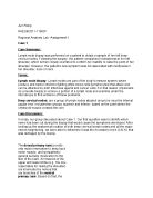
On the contrary, GRK2 inhibition in the adrenal gland and peripheral sympathetic neurons provide sympatholysis. These counteracting effects on the failing heart could be efficient in terms of therapy rather than α2AR agonist treatment. Therefore, the adrenal GRK2 can serve as a specific control of α2AR signaling. Therapy may be focused on this altered relationship between the heart and adrenal gland in HF patients. In general, the data provided by Lymperopoulos et al. suggest treatment for chronic HF can be applied by pharmacological GRK2 inhibitors or targeted GRK2ct gene therapy. Furthermore, these pharmaceutical agents may correct some of the SNS hyperactivity, which leads to HF.20,52
Figure 10: Schematic representation of the pathophysiologic role of GRK2 and the therapeutic potential of its inhibition in heart failure.20
The actual mechanism for GRK2 signaling to the βAR and/or α2AR still remains unknown. There are many postulated mechanisms for these signaling pathways; however an immense amount of research has demonstrated a strong correlation between HF and GRK2 upregulation. However there are many limitations that still need to be taken into consideration, since GRK2 interacts with several intracellular molecules.44 Moreover, GRK2 activity has been observed to inhibit and activate many signaling systems in cardiomyocytes. This has raised many questions regarding its physiological and pathological role in the heart. However, GRK2 inhibition via overexpression of GRK2-ct has shown beneficial effects. Furthermore, GRK2 inhibition has been shown to increase βARs on cells surface. Therefore, GRK2 inhibition may be an interesting therapeutic potential for increased receptor signaling.47 Similarly, GRK2 may also serve as a specific control of α2AR signaling. Therefore, GRK2 inhibition in the adrenal gland and peripheral sympathetic neurons may provide sympatholysis.20 This further produces a counteracting effect on the failing heart which could be efficient in terms of therapy, rather than α2AR agonist treatment. Nevertheless, these studies have shown and provided substantial data and evidence to prove that GRK2 in cardiac and adrenal tissue may be potential pharmaceutical targets in treating HF patients.
In conclusion, despite the modern advances medicine has made in elucidating the mechanisms of heart failure, this disease remains a major cause of mortality in the Western world. It is estimated that in the United States alone, there are 4.6 million patients with heart failure. The prognosis for heart failure is poor, with “median survival after onset only 1.7 years in men and 3.2 years in women.”53 Although there is a tremendous body of experimental knowledge with respect to the changes occurring at the molecular and cellular levels in both hypertrophy and heart failure, it is important at the present time to attempt to tie the seemingly unrelated pathological processes together in order to be able to determine the key triggering process that induces the progression from myocardial hypertrophy to failure. It is important to note, however, that findings at the basic scientific level are not always easily implemented at the clinical level. Thus, even if someday a key cellular trigger for the pathological processes observed in heart failure is found, it might not be possible or feasible to target such a trigger directly with therapy in patients. This knowledge would nevertheless be of great help in devising the appropriate therapeutic strategies that could slow down the progression of heart failure in patients with existing myocardial hypertrophy.
References
-
Petrofski, Jason A. & Koch, Walter J. The β-adrenergic receptor kinase in heart failure. Journal of Moleclar and Cellular Cardiology. 35, 1167-1174 (2003).
-
McMurray JJ & Stewart S. Epidemiology, etiology, and prognosis of heart failure. Heart. 83(5), 596–602 (2000).
-
Gibson TC, White KL, Klainer LM. The prevalence of congestive heart failure in two rural communities. J Chronic Dis. 19(2), 141–52 (1966).
-
Blair AS, Lloyd-Williams F, Mair FS. What do we know about socioeconomic status and congestive heart failure? A review of the literature. J Fam Pract. 51(2), 169 (2002).
-
Cowie MR, Wood DA, Coats AJ, Thompson SG, Suresh V, Poole- Wilson PA, et al. Survival of patients with a new diagnosis of heart failure: a population based study. Heart. 83(5), 505–10 (2000).
-
Lymperopoulos, Anastasios, Rengo, Giuseppe, and Koch, Walter J. Adrenal adrenoreceptors in heart failure: fine-tuning cardiac stimulation. Trends in Molecular Medicine. 13 (12), 503-511 (2007).
-
Leimbach WN, Wallin G, Victor RG, Ayward PE, Sundlof F, Mark AL. Direct evidence from intraneural recordings for increased central sympathetic outflow in patients with heart failure. Circulation. 93, 720–729 (1986).
-
Rockman HA, Koch WJ, Lefkowitz RJ. Seven-transmembrane spanning receptors and heart function. Nature. 415(6868), 206–12 (2002).
-
Cohn, J.N. et al. Plasma norepinephrine as a guide to prognosis in patients with chronic congestive heart failure. N. Engl. J. Med. 311, 819–823 (1984).
-
Port, J.D. and Bristow, M.R. Altered β-adrenergic receptor gene regulation and signaling in chronic heart failure. J. Mol. Cell. Cardiol. 33, 887–905 (2001).
- Keys, J.R. and Koch, W.J. The Adrenergic Pathway and Heart Failure. The Endocrine Society. 13-30 (2004).
-
Caron MG & Lefkowitz RJ. Catecholamine receptors: structure, function and regulation. Recent Prog Horm Res. 48, 277–290 (1993).
-
Brodde OE. Beta-adrenoceptors in cardiac disease. Pharmacol Ther. 60, 405–443 (1993).
-
Bristow MR, Minobe W, Raynolds MV, Port JD, Rasmussen R, Ray PE, Feldman AM. Reduced β1 receptor messenger RNA abundance in the failing human heart. J Clin Invest. 92, 2737–2745 (1993).
-
Inglese J, Freedman NJ, Koch WJ, Lefkowitz RJ. Structure and mechanism of the G protein-coupled receptor kinases. J Biol Chem. 268, 23735–23738 (1993).
-
Eckhart AD, Duncan SJ, Penn RB, Benovis JL, Lefkowitz RJ, Koch WJ. Hybrid transgenic mice reveal in vivo specificity of G protein-coupled receptor kinases in the heart. Circ Res. 86, 43–50 (2000).
-
Harris, Carol A., Chuang, Tsu-Tshen, and Scorer, Carol A. Expression of GRK2 is increased in the left ventricles of cardiomyopathic hamsters. Basic Res Cardiol. 96, 364-368 (2001).
-
Rockman, Howard A., Chiens, Kenneth R., Choi, Dong-Ju, Iaccarino, Guido, Hunter, John. J., Lefkowitz, Robert J., and Koch, Walter J. Expression of a β-adrenergic receptor kinase 1 inhibitor prevents the development of myocardial failure in gene-targeted mice. PNAS. 95, 7000-7005 (1998).
-
Bristow, MR, Ginsburg, R, Minobe, W, et al. Decreased catecholamine sensitivity and -adrenergic-receptor density in failing human hearts. N Engl J Med. 307, 205–211 (1982).
-
Lymperopoulos, Anastasios, Rengo, Giuseppe, Funakoshi, Hajime, Eckhart, Andrea D., Koch, Walter J. Adrenal GRK2 upregulation mediates sympathetic overdrive in heart failure. Nature. 13, 315-323 (2007).
-
Hata, J.A. et al. Genetic manipulation of myocardial b-adrenergic receptor activation and desensitization. J. Mol. Cell. Cardiol. 37, 11–21 (2004).
-
Bylund, D.B. et al. International Union of Pharmacology nomenclature of adrenoceptors. Pharmacol. Rev. 46, 121–136 (1994).
-
Link, R.E. et al. Cardiovascular regulation in mice lacking a2-adrenergic receptor subtypes b and c. Science. 273, 803–805 (1996).
-
MacMillan, L.B. et al. Central hypotensive effects of the alpha2a-adrenergic receptor subtype. Science. 273, 801–803 (1996).
-
Hein, L. et al. Two functionally distinct a2-adrenergic receptors regulate sympathetic neurotransmission. Nature. 402, 181–184 (1999).
-
Brede, M. et al. Feedback inhibition of catecholamine release by two different alpha2-adrenoceptor subtypes prevents progression of heart failure. Circulation. 106, 2491–2496 (2002).
-
Brum, P.C. et al. Abnormal cardiac function associated with sympathetic nervous system hyperactivity in mice. Am. J. Physiol. Heart Circ. Physiol. 283, H1838–H1845 (2002).
-
Small, K.M. et al. Synergistic polymorphisms of b1- and a2C-adrenergic receptors and the risk of congestive heart failure. N. Engl. J. Med. 347, 1135–1142 (2002).
-
Small, K.M. et al. Pharmacology and physiology of human adrenergic receptor polymorphisms. Annu. Rev. Pharmacol. Toxicol. 43, 381–411 (2003).
-
Young, J.B. & Landsberg, L. Catecholamines and the adrenal medulla. in Williams Textbook of Endocrinology 9th edn. (eds. Wilson, J.D., Foster, D.W., Kronenberg, H.M. & Larsen, P.R.) Ch. 17, 665–728 (W.B. Saunders, Philadelphia, 1998).
-
Brede, M. et al. Differential control of adrenal and sympathetic catecholamine release by a2-adrenoceptor subtypes. Mol. Endocrinol. 17, 1640–1646 (2003)
-
Ungerer M, Bohm M, Elce JS, Erdmann E, Lohse MJ. Altered expression of b-adrenergic receptor kinase and b1-adrenergic receptors in the failing human heart. Circulation. 87(2), 454–63 (1993).
-
Ungerer M, Kessebohm K, Kronsbein K, Lohse MJ, Richardt G. Activation of b-adrenergic receptor kinase during myocardial ischemia. Circ Res. 79(3), 455–60 (1996).
-
Gros R, Benovic JL, Tan CM, Feldman RD. G-protein-coupled receptor kinase activity is increased in hypertension. J Clin Invest. 99(9), 2087–93 (1997).
-
Choi DJ, Koch WJ, Hunter JJ, Rockman HA. Mechanism of b-adrenergic receptor desensitization in cardiac hypertrophy is increased b-adrenergic receptor kinase. J Biol Chem. 272(27), 17223–9 (1997).
-
Koch WJ, Rockman HA, Samama P, et al. Cardiac function in mice overexpressing the beta-adrenergic receptor kinase or a beta ARK inhibitor. Science. 268, 1350–1353 (1995).
-
Rockman HA, Ross RS, Harris AN, Knowlton KU, Steinhelper ME, Field LJ, Ross J Jr, Chien KR. Segregation of atrial-specific and inducible expression of an atrial natriuretic factor transgene in an in vivo murine model of cardiac hypertrophy. Proc Natl Acad Sci U S A. 88(18), 8277–8281 (1991).
-
Rockman, H. A., Knowlton, K. U., Ross, J., Jr., and Chien, K. R. Heart Failure: Adaptive and Maladaptive Processes. Circulation. 87, VII-14–VII-21 (1993).
-
Yurenev AP, Parfyonova EV, Krasnikova TL, Aripova NA. Alteration of beta-adrenoreceptor function in hypertensive patients with different degrees of left ventricular hypertrophy. Am J Hypertens. 5,164S-168S (1992).
-
Hunter EG, Hughes V, White J. Cardiomyopathic hamsters, CHF 146 and CHF 147: a preliminary study. Can J Physiol Pharmacol. 62, 1423-1428 (1984).
-
Sethi R, Panagia V, Dhalla KS, Beamish RE, Jasmin G, Dhalla NS. Status of b-adrenergic mechanisms during the development of congestive heart failure in cardiomyopathic hamsters (UM-X7.1). In: Nagano M, Takeda N, Dhalla NS (eds) The Cardiomyopathic Heart Raven Press Ltd. New York, pp 73–86 (1994).
-
Ping P, Anzai T, Gao M, Hammond HK. Adenylyl cyclase and G protein receptor kinase expression during development of heart failure. Am J Physiol 273, H707–H717 (1997).
-
Theilade J., Strom, C. Christiansen, T., Haunso S., and Sheikh, S. P. Differential G protein receptor kinase 2 expression in compensated hypertrophy and heart failure after myocardial infarction in the rat. Basic Res Cardiol. 98:97-103 (2003).
-
Hansen, Jakob Lerche, Theilade Julian, Aplin Mark, and Sheikh Soren P. Role of G-Protein-Coupled Receptor Kinase 2 in the Heart – Do Regulatory Mechanisms Open Novel Therapeutic Perspectives. TCM. 16(5), 169-177 (2006).
-
Ungerer M, Parruti G, Bohm M, et al. Expression of beta-arrestins and betaadrenergic receptor kinases in the failing human heart. Circ Res. 74, 206–213 (1994).
-
Tevaearai HT, Koch WJ. Molecular restoration of beta-adrenergic receptor signaling improves contractile function of failing hearts. Trends Cardiovasc Med. 14, 252–256 (2004).
-
Suzuki, Yoshiyuki, Nakano, Kiyotaka, Sugiyama, Masakazu, Imagawa, Jun-ichi. βARK1 Inhibition Improves Survival in a Mouse Model of Heart Failure Induced by Myocardial Infarction. J Cardiovasc Pharmacol 44(3), 329-334 (2004).
-
Cho MC, Rapacciuolo A, Koch WJ, et al. Defective β-adrenergic receptor signaling precedes the development of dilated cardiomyopathy in transgenic mice with calsequestrin overexpression. J Biol Chem. 274, 17223-17229 (1999).
-
Ishigai Y, Mori T, Moriyama S, et al. Induction of cardiac β-adrenergic receptor kinase 1 in rat heart failure caused by coronary ligation. J Mol Cell Cardiol. 31, 1261-1268 (1999).
-
Maurice JP, Shah AS, Kypson AP, et al. Molecular β-adrenergic signaling abnormalities in failing rabbit hearts after infarction. Am J Physiol. 276, H1853-H1860 (1999).
-
Hoffman, B.B. & Taylor, P. Neurotransmission: the autonomic and somatic motor nervous system. in Goodman & Gilman‘s: The Pharmacological Basis of Therapeutics 10th edn. (eds. Hardman, J.G. & Limbird, L.E.) Ch. 6, 115–154 (McGraw-Hill, New York, 2001).
-
Liggett, Stephen B. Long distance affair with adrenal GRK2 hangs up heart failure. Nature Medicine. 13 (3), 246-248 (2007).
-
Braunwald E, Bristow MR. Congestive heart failure: fifty years of progress. Circulation. 102(20 Suppl 4), IV14-23 (2000).
-
Metaye, Theirry, Gibelin, Helene, Perdrisot, Remy, Kraimps, Jean-Louis. Pathophysiological roles of G-protein-coupled receptor kinases. Cellular Signalling. 17, 917-928 (2005).