Enzyme cofactors
Many enzymes require the presence of an additional, nonprotein, cofactor.
-
Some of these are metal ions such as Zn2+ (the cofactor for carbonic anhydrase), Cu2+, Mn2+, K+, and Na+.
-
Some cofactors are small organic molecules called coenzymes. The B vitamins
are precursors of coenzymes.
Coenzymes may be covalently bound to the protein part (called the apoenzyme) of enzymes as a prosthetic group. Others bind more loosely and, in fact, may bind only transiently to the enzyme as it performs its catalytic act.
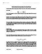
Lysozyme: a model of enzyme action
A number of lysozymes are found in nature; in human tears and egg white, for examples. The enzyme is antibacterial because it degrades the polysaccharide that is found in the cell walls of many bacteria. It does this by catalyzing the insertion of a water molecule at the position indicated by the red arrow. This breaks the chain at that point.
The bacterial polysaccharide consists of long chains of alternating amino sugars:
-
N-acetylglucosamine (NAG)
-
N-acetylmuramic acid (NAM)
These hexose units resemble glucose except for the presence of the side chains containing amino groups.
Lysozyme is a globular protein with a deep cleft across part of its surface. Six hexoses of the substrate fit into this cleft.
-
With so many oxygen atoms in , as many as 14 form between the six amino sugars and certain amino acid such as Arg-114, Asn-37, Asn-44, Trp-62, Trp-63, and Asp-101.
-
Some hydrogen bonds also form with the C=O groups of several .
- In addition, hydrophobic interactions may help hold the substrate in position.
X-ray crystallography has shown that as lysozyme and its substrate unite, each is slightly deformed. The fourth hexose in the chain (ring #4) becomes twisted out of its normal position. This imposes a strain on the C-O bond on the ring-4 side of the oxygen bridge between rings 4 and 5. It is just at this point that the polysaccharide is broken. A molecule of water is inserted between these two hexoses, which breaks the chain. Here, then, is a structural view of what it means to lower activation energy. The energy needed to break this covalent bond is lower now that the atoms connected by the bond have been distorted from their normal position.
As for lysozyme itself, binding of the substrate induces a small (~0.75Å) movement of certain amino acid residues so the cleft closes slightly over its substrate. So the "lock" as well as the "key" changes shape as the two are brought together. (This is sometimes called "induced fit".)
The amino acid residues in the vicinity of rings 4 and 5 provide a plausible mechanism for completing the catalytic act. Residue 35, glutamic acid (Glu-35), is about 3Å from the -O- bridge that is to be broken. The free carboxyl group of glutamic acid is a hydrogen ion donor and available to transfer H+ to the oxygen atom. This would break the already-strained bond between the oxygen atom and the carbon atom of ring 4.
Now having lost an electron, the carbon atom acquires a positive charge. Ionized carbon is normally very unstable, but the attraction of the negatively-charged carboxyl ion of Asp-52 could stabilize it long enough for an -OH ion (from a spontaneously dissociated water molecule) to unite with the carbon. Even at pH 7, water spontaneously dissociates to produce H+ and OH- ions. [] The hydrogen ion (H+) left over can replace that lost by Glu-35.
In either case, the chain is broken, the two fragments separate from the enzyme, and the enzyme is free to attach to a new location on the bacterial cell wall and continue its work of digesting it.
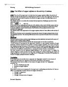
Factors Affecting Enzyme Action
The activity of enzymes is strongly affected by changes in pH and temperature. Each enzyme works best at a certain pH (left graph) and temperature (right graph), its activity decreasing at values above and below that point. This is not surprising considering the importance of
-
(i.e. shape) in enzyme function and
- noncovalent forces, e.g., ionic interactions and hydrogen bonds, in determining that shape.
Examples:
-
the protease pepsin works best as a pH of 1-2 (found in the stomach) while
-
the protease trypsin is inactive at such a low pH but very active at a pH of 8 (found in the small intestine as the bicarbonate of the pancreatic fluid neutralizes the arriving stomach contents). []
Changes in pH alter the state of ionization of charged amino acids (e.g., Asp, Lys) that may play a crucial role in substrate binding and/or the catalytic action itself. Without the unionized -COOH group of Glu-35 and the ionized -COO- of Asp-52, the catalytic action of lysozyme would cease.
Hydrogen bonds are easily disrupted by increasing temperature. This, in turn, may disrupt the shape of the enzyme so that its affinity for its substrate diminishes. The ascending portion of the temperature curve (red arrow in right-hand graph above) reflects the general effect of increasing temperature on the rate of chemical reactions (graph at left). The descending portion of the curve above (blue arrow) reflects the loss of catalytic activity as the enzyme molecules become at high temperatures.
Regulation of Enzyme Activity
Several mechanisms work to make enzyme activity within the cell efficient and well-coordinated.
Anchoring enzymes in membranes
Many enzymes are inserted into cell membranes, for examples,
-
the
-
the membranes of and
- the endoplasmic reticulum
- the nuclear envelope
These are locked into spatial relationships that enable them to interact efficiently.
Inactive precursors
Enzymes, such as proteases, that can attack the cell itself are inhibited while within the cell that synthesizes them. For example, pepsin is synthesized within the (in gastric glands) as an inactive precursor, pepsinogen. Only when exposed to the low pH outside the cell is the inhibiting portion of the molecule removed and active pepsin produced.