Mixings paints and mixing lights bring different results. Paints, like other physical objects reflect certain wavelength and absorb all others, therefore when mixing the colours, the mixture absorb (subtract) more wavelength of light than any of the two does alone. Whilst mixing paints is subtractive, mixing two lights of different wavelength is additive colour mixing. Addictive, as the effect of the wavelengths from each light are added together, thus stimulating more cones. Mixing two paints usually produces a darker colour; in contrast, mixing lights usually produces a lighter colour. In consequence, if more and more different colours of paints are mixed, it results in black, whilst if different coloured lights are added over and over, the combined colour of all wavelengths produces white.
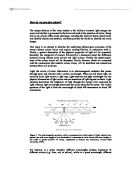
How does the eye covert light energy into neural activity? First the light rays enter the eye from either a natural source (sun) or artificial source (lamp) directly, through an object or as a reflection from an object. The rays travel through the curved, transparent protective layer called the cornea, then passes through the pupil and the also curved lens that is positioned right behind the pupil. The curved surfaces bend the light rays in order to focus them on the surface at the back of the eye, called the retina.
Figure 4. A cross-sectional representation of the eye showing light entering through the pupil. The photosensitive cells, cones and rods, are located in the retina: cones respond to color and rods respond to light intensity (ACEPT W Group, Department of Physics and Astronomy, Arizona State University, 1999).
The retina is an intricate network of cells. Light rays pass through several layers in this network before reaching the photoreceptors. The photoreceptors of the retina code light energy into neural activity via their specialized nerve cells that have photosensitive pigments that respond to light. When rays reach a photopigment, its pigment breaks apart, changing the membrane potential of the receptor cell, thus providing a signal that can be transferred to the brain. The duplexity theory of vision derived from the observation that the retina has two types of photoreceptors: rods and cones. They are not only differing in their shapes, but in their pigments too. The photopigments in rods contains rhodopsin, whereas cones consist of one of three varieties of iodopsin. Rods and cones respond differently to light. Rods are more sensitive to light than cones due to their only one pigment. Therefore rods cannot detect colours, but enable the eye to see even when there is very little light (scotopic vision). On the contrary, cones consist of three different pigments, which become more active at the higher light intensities, therefore enable the eye to distinguish among colours (photopic vision).
Cones and rods also differ in their distribution on the retina. Cones predominate in the area of the sharpest vision, called the fovea, from where rods are absent. The concentration of the cones causes high spatial discrimination, acuity in the fovea, thus facilitating the finely detailed coloured perceptions of the world. With increasing distance from the fovea, the number of cones gradually decreases, and the density of rods gradually increases, demonstrating that rods predominate in peripheral vision. The spatial distribution of rods and cones means that from the central and the peripheral parts of the retina different aspects of vision are perceived. In dim light the more sensitive rods predominate, therefore the produced vision is lack of details and colour perception.
The retina consists of five different layers of cells: receptors, horizontal cells, amacrine cells, and retinal ganglion cells (Pinel, 2003). In order to reach the receptor layer, initially light has to pass through the other four layers. Once the receptor is activated the neural information is transmitted back through the retinal layers to the retinal ganglion cells, whose axons projects across the inside of the retina before forming the optic nerve that expends out of the eye and into the brain. The retinal ganglion cells receive information from a various numbers of cones, and they only become stimulated when the sums of many cones simultaneously activated by light generate an action potential. This explains why retinal ganglion cells that are connected to cones may not respond to dim light, thus colour cannot be detected in faint or dark light conditions. Photoreceptors not only make connections to bipolar cells, but so they do to other types of cells in the retina, including horizontal cells and amacrine cells, which make lateral connections between bipolar cells. The impact of this structural characteristic on vision is that at one point on the retina the response to the light can excite or inhibit the response of a neighbouring cell. For instance, in lateral inhibition the stimulation of one cell is producing a signal that makes it seem as if there is less light at another cell than there really is. The receptive field of a ganglion cell is an area on the retina, to which that cell responds. Most ganglion cells have a centre-surrounding field, and most ganglion cells compare the amount of light stimulating rods and cones in the centre of their receptive field with that stimulating the rods and cones in the surrounding area. Some ganglion cells can only activated by light in the centre of the field and by darkness in the surrounding area, whilst others work in the opposite way; their activity is inhibited by light in the centre of the field and by darkness in the surrounding area. Similarly, the centre and the surround are colour coded; the centre responds best to one colour, and the surround responds best to a different colour. This colour coding is possible as an effect of the varying proportions of the three cone types that feed into the centre and surround of the ganglion cell. (The three types of cones will be discussed later in the essay.) These interactions and processes intensify the sensation of contrast, and influence the sensitivity and acuity of vision.
Three theories have been developed in concerned with the mechanisms that underlie colour vision; the component theory, the contrast theory and the zone theory (Wasserman, 1978). The component theory, also referred to as the trichromatic theory of colour vision considers three spectrally selective component mechanisms in the eye that mediate colour vision. This theory was developed first by Young (1802), and later refined by Helmholtz (1852) based on the experience that any colour can be produced by mixing pure lights of just three wavelengths of light. As there are three different types of cones; each with a different spectral sensitivity, by the combination of the different proportion of excitation in the three elements can produce any colour of the visible spectrum. The three types of cones are the following; the short-wavelength cones detect the shade of blue by responding to the light of about 440 nanometres, whilst the medium-wavelength cones are most sensitive to lights about 530 nanometres (shade of green), finally the long-wavelength cones respond best to light of about 560 nanometres (reddish yellow). Nathans et al. (1992) identified particular genes, which cause this differential sensitivity of cones by directing different cones to produce pigments sensitive to blue, green, or yellow-red. As there is no one single cone that can signal the colour of the light itself, the sensation of a particular colour is based on the ratio of the activities of the three types of cones. Output from many photoreceptors feeds into each ganglion cell, from where the coded information is forwarded towards the brain by the optic nerves.
The contrast theory, also called as the opponent-process theory of colour vision are not describing colour vision as a whole, it only suggest that some antagonistic or opponent processes are involved in the perception of colour. As it was described earlier, the receptive fields of most ganglion cells have a centre field and surrounding area, and they are both colour coded, as altering amount of the three cone types feed into the two different parts of the visual field of a ganglion cell. The centre responds best to one colour, whereas the surround responds best to a different colour. When either the centre or the surround of the ganglion cell is stimulated, the other is inhibited. The colours to which the centre and the surround of a given ganglion cell are most responsive are opponent colours, and the components of each pair inhibit each other. The three pairs described by Hering are red-green, blue-yellow and black-white. Each element signals one colour or the other, but never both. The black-white cells receive information from all types of cones, so it does not matter what colour stimulates them. Stimulating both the centre and the surround cancels the effects of either of both light and produces grey.
Hering’s theory explains two phenomena; on the one hand, certain colours, such as reddish green or yellowish blue never occur, even though it is quite possible to experience bluish green or reddish yellow. On the other hand, this theory explains the colour afterimage phenomenon by proposing that when one component of an opponent pair is no longer stimulated, the other gets automatically activated. Therefore the afterimage of green would be red, blue would be yellow; black’s would be white and vice versa. Hering’s theory is also consistent with the characteristics of the colour circle, as every colour on the circle has a complimentary colour, meaning that when lights of the two colours are mixed together the result is grey. Red and green, yellow and blue, black and white are complimentary colours, which according to the opponent-process theory stimulate the same visual element, but in opposite directions cancelling each other out.
The zone theory is developed on the observation that the visual system is divided into zones. Each of their function is either a component or opponent style; visual receptors function in a component style whereas the central nervous system functions in an opponent mode, therefore together they can explain colour vision.
In summary, the existence of the three types of cones, which has different levels of sensitivities to wavelengths, produces the colour vision according to the trichromatic theory by stimulating them in different ratios. As there are three types of cones, any colour can be produced by mixing three different wavelengths of light. The output of the cones feed into the receptive field of the ganglion cells, where the centre and surround area respond to different colours and inhibit each other. The phenomena of complimentary colours and afterimages are based on these processes. Therefore, the Young-Helmholtz trichromatic theory represents the properties of the photoreceptors; whereas Hering’s opponent-process theory represents the properties of the other layers of the retina. In order to comprehend the complexity of colour perception, both theories need to be taken into account.
References:
ACEPT W Group, Department of Physics and Astronomy, Arizona State University (1999). Color and Light. [Online]. Available: [01/11/2005].
Davies, I. (2005). Lecture notes. [Online]. Available:
[07/11/2005].
Kolb, H., Fernandez, E., & Nelson, R. (2005). Webvision: The perception of color. [Online]. Available: [06/11/2005}
Nathans, J., Merbs, S. L., Sung, C.-H., Weitz, C. J., & Wang, Y. (1992). Molecular genetics of human visual pigments. Annual Review of Genetics, 26, 401-422
Pinel, J. P. J. (2003). Biopsychology. London: Allyn and Bacon.
Sekuler, R., & Blake, R. (2002). Perception. London: McGraw Hill.
Wasserman, G. (1978). Color vision: an historical introduction. Chichester: A Wiley-Interscience Publication.