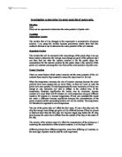
Fig 1: http://faithanatomyg3.wikispaces.com/Intramembranous+Ossification
Endochondral Ossification start on the 2nd month of development, this process involves the mesenchymal to develop into a hyaline Cartilage model which is similar in shape to a bone and that can breakdown, reconstruct and calcify to form a bone. As the bone collar forms the osteoblasts deposit bone which separate from cells and shield the periosteum. The calcification than begins of the cartilage. As the blood vessels enter the midpoint of the diaphysis the primary ossification centre has developed. The ossification centres extend to each epiphysis’s. This allows the bone to elongate and is known as interstitial growth. Secondary ossification centres form at each epiphysis’s and the growth of the bone elongates towards the shaft from each end. (Mosby). The articular cartilage forms from the hyaline cartilage. The hyaline cartilage amid the diaphysis and epiphysis is known as the epiphyseal plate, the epiphyseal plate is only evident before adulthood and is responsible for the growth of the bone. (Tortora p.181)
Fig 2: http://faithanatomyg3.wikispaces.com/Intramembranous+Ossification
The cells found in human bone are extremely important for their formation. As discussed from above the three main cells include osteoblasts, osteocytes and osteoclasts. Osteoblasts; emit unminerialized substances due to their location of regions of high metabolism which leads to them being synthesised, Osteocytes; mature bone cells which regulate the bone mineral content and Osteoclasts which are found on the surface of the majority of adult bones. The two processes discussed above included Intramembranous Ossification which explains the formation of bone in an infant and Endochondral Ossification which explains how the bone forms in cartilage.
B.
Mitosis is the study of the division of the nucleus of a cell which forms two sets of identical sister chromosomes. Mitosis is divided into four phases; prophase, metaphase, anaphase and telophase. However, cell replication also includes the processes known as interphase which is the resting/growth stage and cytokinesis meaning the splitting of the cytoplasm occurs during and after mitosis. Cell aging and death greatly impact the production of cells during mitosis due to errors during DNA replication. (Maireb p.101).
The cycle of mitosis also includes a stage called ‘interphase’ which can also be known as the Growing phase. ’’It is a stage of high metabolic activity’ (Maireb p.91). It is divided into three sub stages; G1 the metabolic activity in the cell is highly active, due to the genetic programming of a cell specific proteins are produced and synthesized while also larger proteins and organelles are manufactured. In the S phase DNA Replication takes place, a set of DNA molecules form. The final phase of interphase G2 ensures the production of enzymes and proteins which are required for the separation and located to the suitable area.(Saladin p.142)
The first phase of Mitosis as mentioned above is prophase, during early prophase chromatin condenses and thickens into chromosomes. The condensing of the chromatin stops the risk of entanglement of the DNA strands. The chromosomes are connected at each centromere and functions to hold the pairs together, the diagram show this process. As the centrioles move to opposite sides of the cell spindle fibres form, positioning themselves on opposing sides. Finally the nucleoli and nuclear membrane recede.(Maireb p.102)
Fig 3:
http://qwickstep.com/search/4-phases-of-mitosis.html
Metaphase is a short process, the diagram shows how the chromosomes have situated themselves along the equator of the cell while also being connected to the spindle fibres at the centromere.(Maireb p.102)
In anaphase the third phase mitosis begins to come apparent due to the breaking of the centromere, the sister chromatids (chromosomes) separate .In the diagram one can see how this separation causes the chromosomes to move towards the opposite poles. The outcome of this process is twice as many chromosomes as previous. (Maireb p.102)
Telophase is the final process of mitosis. From the diagram one can see how the identical DNA molecules (chromosomes) return to their original chromatin state this is done by uncoiling themselves. The nuclear envelope reappears to surround each set of chromosome. Finally the spindle fibres disappear.(Tortora p.92)
Although cytokinesis is not mentioned in the main processes of mitosis it is a crucial process. Cytokinesis is the last process of cell division and means the splitting of the cytoplasm. It usually takes place during or after telophase. The contractile ring appears situated at the cleavage furrows and separates the cell in two (Maireb p.101).
Cell aging is the functional and structural alterations of a cell, causing aging of the cell and in turn on the organism. Therefore cell aging can limit the reproduction of cells. Cell aging is commonly associated with old age. The theory known as ‘wear and tear’, it is believed that the causes of aging include environmental toxins comprising of pesticides, toxins and alcohol which result in errors in DNA replications i.e. Mitosis. Therefore inaccuracies in cells can have severe effects on DNA replication which could lead to mutations within the organisms. In the early development of a cell, death and destruction of the cell are very common. Cell death is known as apoptosis, this process rejects cells that are stressed, aged or cells that are unnecessary. In some cases enzymes are produced which kill the DNA in a cell. Due to cell death, mitosis will not take place if the DNA within the cell has been destroyed. Cell aging and death are crucial processes within the life of a cell, for a good balance of cell reproduction cells need to die so that new stronger cells can reproduce otherwise there would be too many cells. If mitosis was to take place in aging cells mutations of the organism would be a likely outcome. (Maireb p.112).
Mitosis is a very organised and comprehensible process. The four main phases prophase, metaphase, anaphase and telophase describe the division of the nucleus to form two sets of identical sister chromosomes. Interphase describes the growth stage where the cell is preparing for division i.e. producing all the proteins etc. Cytokinesis explains the splitting of cytoplasm which is the final stage of mitosis. Cell aging effects normal cell replication as the cell has been altered functionally and structurally which in turn leads to mutations. However, due to cell death mitosis will not take place in cells if the DNA is destroyed.
References:
Chris Gunn, C.G. (2007). Bones and joints; A guide for students. (5th ed.). London: Churchill-Livingstone.
Patton & Thibodeau,(2010). Anatomy & Physiology. (7th ed.). Missouri: Mosby- Elsevier.
Tortora & Derrickson (2006). Principles of Anatomy and Physiology. (11th ed.). New Jersey: Wiley.
Elaine N. Maireb, E.N.M. (2004). Human Anatomy & Physiology. (6th ed.). San Franciso : Pearson-Benjamin Cummings.
Saladin. (2004). Anatomy and physiology: Unity of form & function. (3rd ed.). New York: McGraw Hill.
:
Fig 2: http://faithanatomyg3.wikispaces.com/Intramembranous+Ossification
http://qwickstep.com/search/4-phases-of-mitosis.html