The basic components of a typical PCR are DNA templates, DNA polymerase, dNTPs, primers, magnesium chloride (MgCl2) and buffer1. A DNA template is needed to act as a base for PCR so that the reaction can occur1. Such template can be man-made or collecting from genomic DNAs, genomic markers, cDNA libraries and RNAs1. DNA polymerase is an enzyme that synthesizes DNA by linking together deoxyribonucleotide monophosphates (dNMPs) in the order detected by the complementary sequence of nucleotides in a template DNA strand8. There are a lot of different types of DNA polymerases, which involves in all sorts of roles but in the case for PCR, mainly polymerases α and β are used8 (table 2 below).
Table 2: Probable roles of some eukaryotic DNA polymerases8
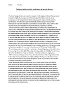
dNTPs are nucleotide triphosphates in which these triphosphates (that comes with the nucleic acid: adenine (A), thymine (T), cytosine (C) and guanine (G)) are found freely in solution and can be assembled together to form oligonucleotide primers in the whole PCR process8. The success of a PCR experiment is also dependent upon the oligonucleotide primers6,8. The primers need to be designed such that one recognizes the sense strand of the DNA to be replaced. Table 3 shows the characteristics of primers while fig 3 shows how an oligonucleotide primer is synthesized chemically:
Table 3: Characteristics of primers6
Fig 3: The synthetic production of oligonucleotide primers6
The production of 5’-AT-3’. Phophoramidite nucleosides are modified with a dimethoxytrityl protecting group on the 5’-end and a β-cyanoethyl protected 3’-phosphite group. Additionally, other modifiers protect primary amines occurring elsewhere in the molecule.
MgCl2 is needed as a cofactor for DNA polymerase while the buffers are required to maintain the environment of the mixture of PCR products8. All these components made up the polymerase chain reaction.
Since stringent conditions and components made up the PCR, such reaction is very specific in building up sequences and uses a lot of different types of amplifications (shown in table 4).
Table 4: Specificity of the PCR reaction2
PCR is advantageous because it is quick and sensitive compared to traditional cloning methods and remains efficient even when the source DNA is heavily degraded or must be isolated from difficult sources such as fixed tissue. However, the cycling tends to be error prone, the size of the products is limited and there is an absolute requirement for prior knowledge of target sequence.
With PCR as the base, there are a lot of advances and extensions to basic PCR strategy. One of them is reverse-transcriptase PCR (RT-PCR), which is the PCR amplification of cDNA2. The first-strand of cDNA synthesis reaction is carried out in a conventional manner but the second strand is synthesized in the first PCR cycle, under the help of thermostable DNA polymerases with reverse transcriptase activity2. RT-PCR is an extremely sensitive method for amplifying the sequences of DNA molecules, and can therefore be used to detect and isolate cDNA sequences from complex sources2. The advantages, disadvantages and uses of RT-PCR are shown in table 5 below:
Table 5: Advantages, disadvantages and applications / uses of RT-PCR2
As a diagnostic technique, RT-PCR can be adapted to both quantity and localized gene products2 (i.e. quantitative and in situ PCRs) (table 6 below).
Table 6: Types of RT-PCR2
In each case, the PCR-based approach is more sensitive than the traditional hybridization-based methods (e.g. RNase protection)2. Building up on this type of PCR, quantitative real-time RT-PCR is introduced6. This type of PCR combines the best attributes of both relative and competitive RT-PCR in that it is accurate, precise, high throughput and relatively easy to perform6. It automates the otherwise laborious process of relative RT-PCR by quantitating reaction products for each sample in every cycle6. Also such system relies upon the detection and quantitation of a fluorescent reporter (STBR® Green and Taqman®), whose signal increases in direct proportion to the amount of PCR product in a reaction6. (Fig 4 shows Taqman® real-time PCR quantification). Other advances of PCR are shown in table 7 below:
Table 7: Other advances of PCR2
Fig 4: Taqman® real-time PCR quantification6
Three primers are used during the PCR process – two of these (primers 1 and 2) dictate the beginning of DNA replication on each DNA strand, and the third (the probe) binds to one strand in between. The probe contains two modified bases – a fluorescent reporter (R) at its 5’-end and a fluorescence quencher (Q) at its 3’-end. As DNA replication proceeds, the extended product from primer 1 dictates the 5’-end of the probe and the exonuclease activity of the polymerase cleaves the fluorescent reporter from the probe. The separation of the reporter from the quencher allows it to fluoresce. The amount of fluorescence is proportional to the amount of PCR product being made and is measured during each PCR cycle.
PCR, as we know, is the most widely used target amplification techniques5. It is most widely applied in in vitro enzymatic amplification but it is not unique2. Several other techniques have developed for particular applications and listed in table 8 below:
Table 8: Alternative in vitro amplification methods2,5
The polymerase chain reaction has revolutionized molecular biology by allowing the amplification and characterization of minute amount of nucleic acids6. As well as being of use to basic scientists, this technique is of immerse importance in medicine for the identification of mutations within small amounts of human DNA, and to pathologists, who routinely need to detect and characterize small amounts of infectious microorganisms6. Other uses of PCR are shown in table 9 below:
Table 9: Applications / uses of PCR1
With the development of more methods, PCR has become much easier and more efficient to obtain more products and used in related or linked molecular biology techniques (i.e. cloning). In the future, it is believed that, together with other methods in molecular biology, PCR will help us to compare the sequence of genes, not only between present-day organisms but also compared to extinct organisms and revolutionized in the world of science and researches8.
References
1 C.R. Newton & A. Graham (1997). PCR. BIOS Scientific Publishers Ltd.
2 R.M. Twyman (1998). Advanced molecular biology. BIOS Scientific Publishers Ltd
3 R.K. Saiki, S. Scharf, F. Faloona et al (1985). Enzymatic amplification of beta-globin genomic
sequences and restriction site analysis for diagnosis of sickle cell anemia. Science. 230: 1350-
1354.
4 K. Mullis, F. Faloona, S. Scharf, R. Saiki, G. Horn & H. Erlich (1986). Specific enzymatic
amplification of DNA in vitro: the polymerase system. Cold Spring Harbor Symp Quant Biol.
51: 263-273.
5 A.E.M. Salvatore, T. Sanjay & R.K. Fred (2006). Real-time Assays with Molecular Beacons
and Other Fluorescent Nucleic Acid Hybridisation Probes. Clinica Chimica Acta. 363: 48-60.
6 J.R. Richard (2004). Analysis of genes and genomes. John Wiley & Sons Ltd.
7 H. Stokes (2007). CBMS 352 molecular biology laboratory manual. Macquerie University,
Department of Chemistry and Biomolecular Sciences.
8 F.W. Robert (2005). Molecular Biology (International edition) (3rd edition). McGraw Hill
companies.
9 B. Janet (2006). Applications of real-time immuno-polymerase chain reaction (rt-IPCR) for
the rapid diagnoses of viral antigens and pathologic protein. Molecular Aspects of Medicine.
27: 224–253.
10 F. Watzinger, K. Ebner & T. Lion (2006). Detection and monitoring of virus infections by
real-time PCR. Molecular Aspects of Medicine. 27: 254–298.
11 T.S. Joanne (2006). Real-time PCR for prenatal and preimplantation genetic diagnosis of
monogenic diseases. Molecular Aspects of Medicine. 27: 176–191.
12 A.B. Stephen & M.R. Mueller (2006). Real-time reverse transcription PCR and the detection
of occult disease in colorectal cancer. Molecular Aspects of Medicine. 27: 192-223.