Aetiology
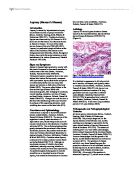
Leprosy is a chronic granulomatous disease caused by the acid-fast bacillus Mycobacterium leprae (Deepa & Ebenezer 1999:83.173). (Figure 1).
Figure 1: Rod shaped bacillus that causes leprosy.
It is identical in appearance to M. tuberculosis and is aerobic; rod shaped, and typically stains in a beaded manner (Nester, Anderson, Roberts, Pearsall & Nester 2004:671). M. leprae is an exceptional bacterium because of its long generation time and no growth in artificial media (Sasaki, Takeshita, Okuda & Ishii 2001:45.729); its generation time is about 12 days (Nester, Anderson, Roberts, Pearsall & Nester 2004:671). It also has a long incubation period of 2-5 years (Oskam 2004).
Pathogenesis and Pathophysiological Features
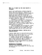
M. leprae grows intracellularly (Mims, Dockrell, Goering, Roitt, Wakelin & Zuckerman 2004:358). The principle target cell for the leprosy bacillus is the Schwann cell, and the resulting nerve damage is responsible for the main clinical features of leprosy (Greenwood, Slack & Peutherer 1997:210). The bacterium initially grows very well within macrophages, before delayed hypersensitivity develops (Nester, Anderson, Roberts, Pearsall & Nester 2004:671). Immune macrophages limit the growth of M. leprae, and the bacteria therefore do not become numerous (671). The spectrum of disease activity is very broad depending upon the presence or absence of a cell-mediated immune (CMI) response to M. leprae (Mims, Dockrell, Goering, Roitt, Wakelin & Zuckerman 2004:358). At one end of the spectrum is tuberculoid leprosy (TT), which carries a better prognosis than lepromatous leprosy (LL). In some patients TT is self-limiting, but in others it may progress across the spectrum towards LL (358). The features of lepromatous leprosy are skin lesions causing nodules (figure 2) and diffuse thickening, and the characteristic ‘leonine facies’ (Timbury, McCartney, Thakker & Ward 2003:230). As the disease progresses there is loss of eyebrows, thickening and enlargement of the nostrils, ears and cheeks (Mims, Dockrell, Goering, Roitt, Wakelin & Zuckerman 2004:358). The mucus of the nose and throat is loaded with the bacteria, which can readily be transmitted to others (Nester, Anderson, Roberts, Pearsall & Nester 2004:671)
Figure 2: A man with nodules on his face as the result of Lepromatous leprosy. With treatment this disfigurement can be almost entirely reversed.
Tuberculoid leprosy shows lesions that are localised to the skin and peripheral nerves, with anaesthesia (Timbury, McCartney, Thakker & Ward 2003:230). Repeated injury to, and infections of, the anaesthetic extremities leads to their gradual destruction (Greenwood, Slack & Peutherer 1997:210). The nasal bones and their destruction may lead to collapse of the nose (211). The eye is frequently damaged by direct bacillary invasion, uveitis or corneal infection secondary to paralysis of the eyelids (211). People with tuberculoid leprosy rarely, if ever, transmit the disease to others (Nester, Anderson, Roberts, Pearsall & Nester 2004:671).
Prognosis and Treatment
If the disease is diagnosed early and treatment initiated promptly the patient has a much better prognosis (Mims, Dockrell, Goering, Roitt, Wakelin & Zuckerman 2004:359). The multi-drug treatment (MDT) is considered to be the best way to treat leprosy today. Tuberculoid leprosy can be arrested by treatment with the antimicrobials dapsone and rifampin administered in combination for six months (Nester, Anderson, Roberts, Pearsall & Nester 2004:672), and has a better prognosis than lepromatous leprosy (Mims, Dockrell, Goering, Roitt, Wakelin & Zuckerman 2004:358). Lepromatous leprosy is treated with dapsone, rifampin and clofazimine and is given for a minimum of 2 years and may be lifelong or until all skin scrapings and biopsies are negative for acid-fast rods (358). It is often necessary to correct deformities, prevent blindness and further damage to anaesthetic extremities, and treat reactions with anti-inflammatory drugs and to attend to the patient’s social, psychological and spiritual welfare (Greenwood, Slack & Peutherer 1997:212).
Glossary
Granuloma: Found in a chronic inflammatory response, collections of lymphocytes and stages of macrophages; an attempt by the body to wall off and contain persistent organisms and antigens. Incubation period: The time it takes for symptoms to develop after initial exposure to the disease-causing organism. Leonine: Lion like. Lesions: Any visible, local abnormality of the tissues of the skin. Nodules: A small mass of tissue in the form of a protuberance or a knot that is solid and can be detected by touch. Uveitis: Inflammation of the uvule tract, layer of tissue in the eye comprises the iris and its supporting structures and the choroid
membrane behind the retina.
References
* Cole et al (2001) Massive Gene Decay in the Leprosy Bacillus. Nature 409, 1007-1011.
* Deepa J & Ebenezer D (1999), Infectious Keratinitis in Leprosy. Br J Opthalmol 83, 173-176.
* Greenwood D, Slack R & Peutherer J (1997) Medical Microbiology (15th edition). Pearson Professional Ltd.
* Katoch V (2002) Advances in the Diagnosis and Treatment of Leprosy. Expert Reviews, 77, 1-8. (accessed 24/02/2004).
* Mims C, Dockrell H, Goering R, Roitt I, Wakelin D & Zuckerman M (2004) Medical Micrcbiology (3rd edition). Mosby-Year Book Europe Ltd.
* Nester E, Anderson D, Roberts C, Pearsall N & Nester M (2004) Microbiology a Human
Perspective (4th edition). Vann Hoffmann Press, Inc.
* Oskam L (2004) Leprosy. (accessed 24/02/2004)
* Pataki G (2003), Leprosy.
* Sasaki S, Takeshita F, Okuda K & Ishii N (2001) Mycrobacterium Leprae and Leprosy: A Compendium. Microbiol. Immunol. 45, 729-736.
* Timbury M, McCartney A, Thakker B & Ward K (2003) Notes on Medical Microbiology. Elsevier Science Ltd.