As the path of the electron beam could be impeded by collisions between the electrons and air particles, the electron beam passes through a vacuum. As there is a vacuum, only dead specimens can be examined. Also the specimen preparation routine requires the specimen to be dead anyway.
With a TEM, a permanent record of the image can be taken, due to the presence of a photographic plate. The image produced is called an electron micrograph. The electron beam can also be reflected into a viewing port. The fluorescent screen, which reflects the electron beam, is coated with electron sensitive compounds, as the image produced by the electron beam cannot be viewed directly. It needs to be converted into a format which can be viewed by human eyes.
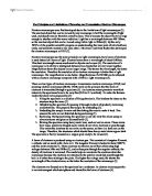
Specimen Preparation
Fixed
↓
Dehydrated
↓
Cleared
↓
Embedded
↓
Sectioned
↓
Stained
↓
Mounted
The stages in preparation of the specimen for viewing by a TEM are as above.
The specimen is firstly Fixed to avoid distortion of cell components. The tissue is killed, and then preserved in as natural a state as possible by a chemical fixative. This works by denaturing the cells constituent protein. In electron microscopy, glutaraldehyde is used as the chemical fixative.
The specimen is then Dehydrated. This process is gradual in order to preserve fine detail. This is done by using a series of progressively increasing concentrations of either ethanol or propanone.
The sample is then Cleared. This is done because the alcohol may be immiscible with embedding agents, and is thus replaced by a clearing agent that also has the effect of making the material transparent. The most commonly used cleaning agent is xylol.
The material is then Embedded in plastic or resin, to provide support during sectioning. This prevents deformation of the specimen during the process of sectioning.
Sectioning is the process of cutting the material into extremely thin sections for viewing by the electron microscope. The ultra thin sections, of thickness 20-100 nm are cut using a diamond or glass knife. The machine used is called am ultramicrotome. The material must be cut into sections because the electron beam has a low penetrating power.
The sections produced are then Stained with solutions of heavy metal salts in order to improve contrast. This works because biological structures are almost transparent to electrons, and the contrast is improved by increasing the number of electrons that are deflected by the sample. The presence of heavy metal salts, such as lead acetate will increase the number of deflected electrons, and thus improve the contrast of the resulting image.
Finally, the specimen is Mounted on a thin copper grid, measuring 3mm in diameter. This is used because electron beams cannot pass through glass, yet the electrons can pass through the gaps present in the grid.
Although images produce by the TEM have a very high resolution, and can have high magnification, several electron micrographs may be required to give a composite image of the specimen. Also as ultra thin sections of a sample are analysed, a number of the said sections are required to provide a 3d image of the sample.
Scanning Electron Microscope (SEM)
The scanning electron microscope works in a similar way to the TEM. An electron beam is fired by an electron gun, again part of the cathode. The electron beam passes through two electromagnetic lenses prior to striking the specimen which focus the beam, and magnify the image. However as the electrons do not pass through the specimen, images produced by a SEM are not produced by the primary electrons from the electron beam. What actually happens is that the primary electron beam knocks electrons from the surface of the sample. These secondary electrons are then picked up by a collector, then amplified, prior to being transmitted to a viewing screen, or a photographic plate to produce an electron micrograph. Because the image is produced from secondary electrons, both the resolution and the magnification are less than is possible with a TEM, 10 nm, and 100x. However, the SEM provides a true 3d image of the external appearance of a sample, and can take larger samples. It can also provide a 3d image of the inside of a specimen via a technique called freeze fracturing.
Freeze Fracturing is a technique whereby a sample is frozen in liquid nitrogen, and then split open. It allows interior detail to be seen with a SEM.
Cells are frozen at -196°C and then split. The exposed surfaces are then coated with carbon and platinum. The organic material is then dissolved with enzymes by a process called freeze etching, leaving a carbon-platinum replica of the original surface, which can be examined with the microscope.
As with the TEM, there is a preparation procedure for the usage of samples with the SEM. However, it is much less complex than the TEM procedure. Samples merely have to be coated with a thin film of carbon or gold.
In conclusion, both types of electron microscope have advantages and disadvantages. The TEM has a high magnification and resolution compared to the SEM, but the SEM can give a true 3d image from one electron micrograph. Thus, the best type of microscope to use for any given sample would depend on the sample, and the sort of image that is required. For completion, both the TEM, and the SEM would have to be used in conjunction with each other. The advantages of one complement the advantages of the other.