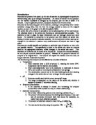
- Enzymes usually work within a very narrow pH range.
-
The range is dependant on the nature of the active site; whether it accepts or donates H+ ions when binding.
- If the substrate is always in excess then increasing the enzyme concentration will increase the rate, in a linear relationship.
-
Not a linear relationship, gives a curve reaching a plateau (Vmax )
- The amount of enzyme becomes the limiting factor because it has a set rate of conversion (turnover rate) that cannot be exceeded, no matter how much substrate is present
-
The amount of substrate needed to reach ½Vmax is called Km.
-
The rate can be found by using the equation:
Aims
The aim of this investigation is to study the effect of pH on the catalytic activity of the enzyme pepsin and to determine it’s optimum working pH.
We plan to do this by measuring the amount of time it takes for pepsin to break down the gelatin-binding layer on a strip of camera film in a series of buffer solutions.
Hypothesis
Pepsin will degrade the gelatin-binding layer more quickly at pH4, as this is the pH that pepsin works at in the stomach, so this must be the optimum pH.
Apparatus
6 boiling tubes
Water bath
1% Pepsin solution
pH buffers 1, 2, 3, 4, 5, 6
Stop clock
Photographic film (Ilford No4)
Scissors/craft knife and ruler for cutting film
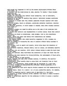
Method
- 2ml of 1% pepsin solution was added to 6 boiling tubes.
- To each boiling tube 2ml of a pH buffer was added. So each tube had a different buffer (1-6).
-
The boiling tubes were then placed into a water bath at 45oC. The tubes were then left to equilibrate for five minutes.
- 1 cm² of photographic film was then placed into each boiling tube and the stop clock started.
- The visual appearance of the photographic film in each boiling tube was monitored and the time recorded when and if the photographic film in each boiling tube turned translucent.
- To improve accuracy an average time was obtained for each pH buffer solution by the repetition of the above method.
Risk Assessment
In this investigation there are a number of risks to safety:
A first aid kit, including eyewash should be kept to hand at all times.
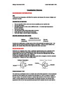
Results
This computer drawn graph shows that the optimum pH is just below 3, however this was not determined experimentally. From our results we can only say that the optimum pH was 3, the hand drawn graph (over) reflects this.
Discussion
The base of 35mm photographic film is celluloid and there are many layers on top of this. The most important layer is the photosensitive silver halides that react with light to produce the picture. Gelatin is a protein extracted from animal hides and used to suspend the light sensitive silver halides and bind them to the celluloid base.
Pepsin is the main protease found in the stomach of mammals and other animals. It digests proteins into smaller, peptide molecules, which can be readily absorbed by the intestinal lining. Pepsin is synthesized in an inactive form by the stomach lining; hydrochloric acid, also produced by the gastric mucosa, is necessary to convert the inactive enzyme and to maintain the optimum acidity for pepsin function.
When the photographic film is dropped into the boiling tube, the pepsin begins to break down the gelatin and the silver halides are removed from the celluloid layer making the film transparent.
The pH of a solution can have several effects of the structure and activity of enzymes.
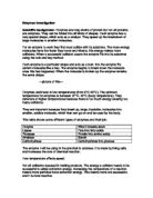
For example, pH can have an effect of the state of ionisation of acidic or basic amino acids. Acidic amino acids have carboxyl functional groups in their side chains. Basic amino acids have amine functional groups in their side chains. If the state of ionisation of amino acids in a protein is altered then the ionic bonds that help to determine the 3-D shape of the protein can be altered. This can lead to altered protein recognition or the enzyme might become inactive. This is similar to what happens with pepsin; Pepsinogen contains an amino-terminal precursor segment of 44 residues that is proteolytically removed in the formation of Pepsin. This activation normally occurs below pH5. Cleavage of the peptide bond between leucine 16 and isoleucine 17 in the precursor segment is absolutely essential.
X-ray crystallographic analyses show that the active site is fully formed in pepsinogen but is blocked at neutral pH by residues of the precursor segment. Salt bridges are formed by six lysine and arginine side chains with carboxylate side chains of glutamate and asparate residues of the pepsin moiety when several carboxylates are protenated the pH is lowered, salt bridges between the precursor segment and the pepsin moiety are then broken. The peptide bond is now hydrolysed between the precursor and pepsin moieties (www.lancs.ac.uk, 2000). Both the structure of pepsin (top), and pepsinogen (bottom) can be seen to the left (protein data bank, -).
In general enzyme have an optimum working pH. However the optimum is not the same for each enzyme.
The pH in the stomach can vary greatly between pH1 and pH5 or more, so it would be acceptable to think that pepsin would work between these pHs. However, pepsin works in a much narrower pH range, from the table and graph we can see that the pepsin worked most effectively at pH3, and worked very slowly in only three other buffers, therefore between somewhere between ph2 and pH4 is pepsin’s optimum. This is probably closer to pH 2 as it worked faster there than at pH 4.
Further Work
To improve on this investigation we could carry out some further experiments.
- Intermediate pHs should be tested to determine the exact optimum pH.
- The effect of temperature should be tested so we could find the perfect conditions for pepsin.
- Varying the substrate and enzyme concentrations would also give us an idea of the perfect working conditions for pepsin.
- There were a number of errors in our investigation that should be thought about:
- The buffers may not have been the exact pH stated.
- The pepsin solution may not have been exactly 1%.
- The film may not have been cut exactly to size.
- The temperature of the water bath fluctuated slightly.
- The amount of agitation to each tube may not have been the same.