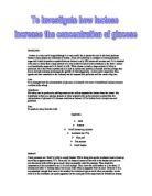
Glucose two different structures.
6CH2OH
H C O H
H
C OH H C
OH C C OH
H OH
Alpha glucose
6CH2OH
H C O OH
H
C OH H C
OH C C H
H OH
The differences is, on the alpha glucose structure the possition of H and OH on the right hand side of the structure is different to the position there are in beta glucose.
As we know that glucose are sugars, so there are different types of them. Examples are
- Maltose, which is found in malt and it’s made up of 2 glucoses.
- Sucrose, it’s normally found in table sugar and it’s made up of glucose and fructose. (Fructose is another source of sugar)
- Lactose, it’s normally found in milk and it’s made up of glucose and galactose.
All these sugar are disaccharides, which means, two simple sugars. Before they disaccharides they are monosaccarides the disaccarides are made by condensation process. If wanted to change the disaccharides back to monosaccarides hydrolysis process is used.
When eat too much sugar we have a blood sugar problem or medical term Diabetics. The test used
The test for glucose is in two ways; one is semi quantitative method and secondly is Quantitative methods. The both methods involve using benedict reagent.
Quantities benedict’s is tested by mixing volume amount of solution desire to test with equal volume amount of Benedict’s reagent. Then the solutions are heated gently, while heating colour precipitated gently appears, which shows the present of sugar or glucose in the solution.
The colour of the precipitate changes from yellow to orange to red etc., this shows how high or low of concentration there is in the solution. This test can also be used to test for blood sugar level. While same sort of result could be observed.
Aim: How to use quantity of glucose concentration in body fluid in clinical diagnosis.
Outline methods: In this experiment to find out if a substances contain glucose both semi quantitative and quantitative benedict’s test can be done. This test is also used for blood sugar test in clinical diagnosis.
But in this case quantitative Benedict’s test procedures is used.
Solution is added with benedict’s reagent and it’s placed in water bath, then it’s centrifuged to settle the participates solution at the bottom, while the clear solution at the top. The clear solution is then poured in cuvette to read in spectrophotometer. And the reading are recorded in Optical Density (OD)
Key Controlled Variables: In this experiment some things will have to be kept variable. These are
-
Volume of water which should be 3cm3
-
Volume of glucose, which should be 3cm3 , but at different concentration in each test tube.
- Drops of Benedict’s reagent, which are 4.
Keeping all these will give more chance of getting a reliable result.
Equipment
- 6 clean Test Tubes
- 1 Test Rack
- 6-7 Pipette
- Water Bath
- Spectrophotometer
- Centrifuge
- Centrifuge Tubes
- Cuvette
Ingredient
- 5% concentration of glucose
- Distilled Water
- Benedict’s Reagent.
Procedures
-
Collect 6 clean test tubes (sizes doesn’t matter)
- Collect I test tube rack.
- Position the 6 test tubes in test tube rack
- Labelle the test tubes letter A-F (alphabetically)
-
Pipette 6cm3 of 5% glucose concentration in test tube a.
-
Using clean pipette, pipette 3cm3 of distilled water in test tube B to F
-
Using clean pipette, take 3cm3 of 5% glucose concentration from test tube A, then add to test tube B to make the concentration of glucose in test tube B 2.5%
-
Using clean pipette, pipette 3cm3 of solution in test tube B to test tube C to make the concentration of glucose 1.5%
-
Using clean pipette, pipette 3cm3 of solution in test tube C, then add to test tube D to make the concentration of glucose solution 0.625%
-
Using clean pipette, pipette 3cm3 solution from test tube D, then add to test tube E to make glucose concentration in test tube E 0.312%
-
Using clean pipette, pipette 3cm3 solution from test tube E, then waste it. Wasting the solution make Test tube F left with just distilled water. In another word 0% of glucose.
- Add 4 drops of benedicts to each solution in every test tube (A-F), and then mix the two solutions together.
- Place the mixed solution of in water bath for few minutes. (Cover the water bath to make the reaction quicker).
- After few minutes take the solution out of the water bath, while doing this “care” because of hot surface.
- Record in a table the colour of each solution alphabetically.
- Pour each solution alphabetically in centrifuge tube. Labelle centrifuge tube 1,2,3 instead of A, B, C, and then centrifuge the solution.
- After centrifuging the solutions, pour them in cuvette in order to read in spectrophotometer.
- When reading on spectrophotometer display –ve, it means +ve, but the +ve could be invisible.
- Record the result of spectrophotometer in unit of optical density (OD) on a table.
- Repeat the procedure to observe more reliable result.
This is the exact procedure that could be used to test for concentration of sugar in body fluid.
Result table.
Graph: On graph sheet
Conclusion. The graph shows result of quantitative Benedict’s test, which is concentration against spectrophotometer reading. The two reading were added together then divided by two to get the average reading, so there fore average reading is plotted on the graph to show the result. Unfortunately it shows on the graph that the result isn’t accurate. When look at the graph it shows that the reading of spectrophotometer changes while they should be dropping accurately due to the fact that the concentration of glucose is dropping accurately. I.e. The concentration of glucose is high, so is the reading should be high. Unfortunaly on the graph when the concentration of glucose was at 5% the average reading was .193 and when concentration drop to 2.5% average reading gives was .363, which is higher than what it should be.
Evaluation. The in investigation on glucose, and using quantitative benedict’s test to observe the presence of glucose in solution was quite understandable also the procedure used, but unfortunately the result was no where being accurate. This is because where the reading meant to be high, because the concentration is wasn’t.
The inaccurate results are understood to be anomalous result.
Thinking of what might have make the result inaccurate might have something to do with the procedure used. To make the result more reliable, it would be recommendable that the method should be repeated with a lot of care.
As aimed is concerned, which is using this procedure in clinical diagnosis, this won’t be possible, because unreliable the result is. But to make it reliable a very considered, clean, care, and imaginative procedure could be used to make a firm conclusion on how to use the quantitative test for clinical diagnosis, which could give a reliable result.
Medically these will discovered how high blood sugar concentration patient have, which is by examining his or her fluid (urine).
Other way to test for blood sugar level is by using a method called glucose tolerance test. This when doctor ask patient to swallows a sugar solution, then the doctor measure the blood glucose concentration at interval.