Aim:
To observe the different stages of mitosis, of an onion root tip.
Equipment:
- Onion
- Tweezers
- Clamps
- Compound light microscope
- Slides
- Cover slip
- Hot plate
- Acidified Orecin Stain
- Watch glass
- Stop watch
- Drawing kit
Method:
- Prepare an onion by placing it in water for ten days.
- Cut the onion into small parts using sharp scalpel.
- Tear a thin piece of tissue from the epidermal layer from the inner side of the slice using clamps. Notice that the epidermal layer is a thin transparent layer, which covers the leave.
- Place the thin tissue on the clean slide. Cut a (2x3mm) portion of the epidermal with a sharp scalpel. Avoid any folding to the epidermal layer.
- This step is related to the previous step, because it will show the proper way to stain a slide:
- Place one drop of Acidified Oricen stain on one edge of the cover slip, and the flat edge of a piece of paper towel on the other edge of the cover slip. The paper towel will draw the water out from under the cover slip, and the cohesion of the water (due to those perennial favorites - Hydrogen Bonds).
- As soon as the stain has covered the area containing the specimen you are finished. The stain does not need to be under the entire cover slip. If the stain does not cover the area needed, get a new piece of paper towel and add more stain until it does.
- Be sure to wipe off the excess stain with a paper towel, so you don’t end up staining the objective lenses.
-
In the meanwhile place the watch glass on the hot plate at (500C) for about two minutes.
- Use a compound light microscope to examine your wet preparation under low power, and then use the high power magnifications.
- As soonest as you get a clear image of the of the onion cell, observe the development stages and draw each stage as you see them under the microscope.
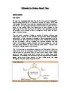
Results:
The following picture show mitosis in onion root tips as it was observed under the compound light microscope:
Discussion of results:
Mitosis is the mechanism that allows the nuclei of cells to split and provide each daughter cell with a complete set of chromosomes during cellular division. This, coupled with cytokinesis (division of the cytoplasm), occurs in all multicellular plants and animals to permit growth of the organism. In this experiment, we observed the various steps in mitosis that occur in onion root tips.
Interphase is the stage in which the chromatin is undifferentiated in the heavily stained nucleus. Before the cell enters the mitosis phase, it first undergoes a synthesis or S-phase where each chromosome is duplicated and consists of two sister chromatids joined together by a specific DNA sequence known as a centromere. Centromeres are crucial to segregation of the daughter chromatids during mitosis.
The first phase of mitosis is known as the prophase, where the nuclear chromatin starts to become organized and condenses into thick strands that eventually become chromosomes. During prophase, the cytoskeleton (composed of cytoplasmic microtubules) begins to disassemble and the main component of the mitotic procedure, the mitotic spindle begins to form outside the nucleus at opposite ends of the cell.
The next identifiable phase is called metaphase where the chromosomes, attached to the kinetochore microtubules, begin to align in one plane (the metaphase plate) halfway between the spindle poles. The kinetochore microtubules exert tension on the chromosomes and the entire spindle-chromosome complex is now ready for the next event.
This sets the stage for chromosome separation in the next stage of mitosis is anaphase. Almost immediately after the metaphase chromosomes are aligned at the metaphase plate, the two halves of each chromosome are pulled apart by the spindle procedure and migrate to the opposite spindle poles. The kinetochore microtubules shorten as the chromosomes are pulled toward the poles, while the polar microtubules elongate to assist in the separation.
In telophase, the daughter chromosomes arrive at the spindle poles and are eventually redistributed into chromatin. The process of cytokinesis, where the cytoplasm is divided by cleavage, also starts sometime in anaphase and continues through telophase. After complete separation of the chromosomes and their extrusion to the spindle poles, the nuclear membrane begins to reform around each group of chromosomes at the opposite ends of the cell. The nucleoli also reappear in what will eventually become the two new cell nuclei.
When telophase is complete and the new cell membrane (or wall in the case of the onion root tips) is being formed, the nuclei have almost matured to the pre-mitotic state. The final steps are completion of the total formation of a membrane between each of the new daughter cells to yield two separate new cells.