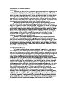
Fusidic Acid (dark green0
Methicillin (gold)
Novobiocin (lilac)
Penicillin G (Pink)
Streptomycin (Blue)
Tetracycline (Brown)
Hypothesis
Some of the antibiotics will have an effect on the bacterium S. albus.
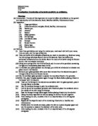
Method
Ensure that safety goggles and a lab coat are worn.
Using the aseptic technique 3 petri dishes are set up individually. (See Diagram 2)
Before starting disinfect the work surface to avoid contamination.
Experimental Dish 1
Step 1
Using a sterile pipette, transfer 1cm of a broth culture of Staphylococcus albus into a sterile Petri-dish and replace the lid immediately. Ensure that the neck of the bottle containing the S albus is placed in a blue Bunsen burner flame before and after taking the sample.
Step 2
Add approximately 25cm of sterile nutrient agar using another sterile pipette and replace the lid immediately.
Step 3
Very gently move the petri-dish side to side, up and down and in circles to spread the bacterial cells throughout the agar. Allow the nutrient agar to cool and set.
Step 4
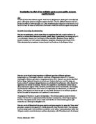
Once set, using a sterile pair of forceps, place the Mastring disk onto the set agar and replace the lid. The Mastring disk is a paper disk with 8 coloured disks attached to it.
These 8 disks are impregnated with a known antibiotic. (See Picture 1 below)
Picture 1 Photo of Mastring being used in Antibiotic Sensitivity Test
Control Dish 2
An identical dish was set up using steps 1-3 as above. This will be a control dish to compare S. albus growth with and without antibiotics present.
Control Dish 3
A third dish containing 25cm of sterile nutrient agar only was set up also as a control.
This control will check that the agar was sterile and that the aseptic technique was carried out correctly.
Once the agar had set in all 3 petri-dishes, seal with tape and turn upside down to prevent condensation forming on the plates and incubated for 48 hours at 25C.
Disinfect work surface again and was hands thoroughly.
Results
After incubation the petri-dishes were observed, remembering not to open the dishes.
There may be pathogenic organisms present in the dishes which could be harmful.
S. albus is not harmful but it may have mutated during the experiment.
The experimental dish showed varying degrees of inhibition of the bacterium by some of the antibiotics indicated by a clear ring. (See Diagram 3)
The effect of the antibiotics was measured in mm as shown in the Table of Results below.
Table of Results
ANTIBIOTIC AREA OF CLEARZONE (Unit mm )
-----------------------------------------------------------------------------------------------
Chloramphenicol (green) 355mm
Erythromycin (red)
Fusidic acid (dark green) 1230mm combined (overlap)
Methicillin (gold)
Novobiocin (lilac) 0mm
Penicillin G (pink) 27mm
Streptomycin (yellow) 115mm
Tetracycline (brown) 521mm
The results are also illustrated in the following bar chart. (Diagram 4)
Discussion
The experiment was worthwhile as it could identify the most effective antibiotic to treat a particular bacterial infection assuming that the agar and bacteria were mixed properly and that there was no contamination.
Conclusion
The most effective antibiotic to inhibit S. albus was Tetracycline as the clear area was very well defined.
Some of the other antibiotics were not as effective, the clear area was not so well defined and in some cases the areas overlapped and made it difficult to measure there effect accurately. The result did support the hypothesis.
Control Dish 2 was cloudy in appearance, which indicated a uniform growth of
S. albus, throughout the agar. This suggests that the agar had been prepared correctly to enable the growth of the bacteria. (See diagram 5)
Control Dish 3 was completely clear, which indicated no growth of micro organisms and that the aseptic technique was carried out correctly. (See diagram 5)
Further Work
A different bacterium could be used to observe whether the same antibiotic was as effective as on the S. albus.
The same bacteria could be used but isolated with just one antibiotic to get a clearer result.