-
Cohesin breakdown is caused by a called separin (also known as separase).
- Separin is kept inactive until late metaphase by another protein called securin.
-
Anaphase begins when the destroys securin (by tagging it for deposit in a ) thus ending its inhibition of separin and allowing separin to break down the cohesins.
4. Telophase
A nuclear envelope reforms around each cluster of chromosomes and these return to their more extended form.
Phosphate is important for all organisms; it is essential for growth and development and has several roles;
- Nucleotides – DNA, RNA & ATP are nucleic acids; phosphate groups are vital to their structure. It combines with the sugar forming a sugar-phosphate backbone, which holds the molecule and covalently bonds different nucleotides to one another. DNA & RNA have one phosphate group and ATP has three. ATP is used by cells to carry out ‘work, therefore is considered as ‘energy currency’ and is needed during the actual process of mitosis.
- DNA replication - two extra phosphate groups are added to nucleotides for activation, allowing them to pair up with complementary bases on the ‘old’ DNA strands (semi-conservative replication).
- Protein synthesis - phosphate groups are required during transcription to produce mRNA otherwise protein synthesis can’t occur; thus no proteins will be made.
- Phospholipids make up the plasma membrane, the phosphate group makes the head hydrophillic whilst the fatty acid tails remain hydrophobic, this is very significant for the membrane’s characteristics. New membranes are synthesised (cytokinesis), therefore phosphate must be available.
( I have obtained some of the factual information above this line from my school notes and my OCR biology 1 textbook)
Experiment Prediction:
High levels of phosphate allows more ATP to be produced so more energy is available. Mitosis is an ‘ active’ process, ATP will be needed by the energy consuming cells. Needed to activate nucleotides in cytoplasm so DNA can be replicated. Insufficient amounts of phosphate means that DNA nucleotides can’t be made or activated, therefore no replication. More phosphate ions allows more nucleotides (DNA & RNA) to be produced, as they are a component of the backbone. Daughter nuclei produced will go through cytokinesis and phosphate ions will be needed to synthesise new membranes. Lack of phosphate means protein synthesis won’t occur or will be inefficient, vital enzymes needed during mitosis can’t be made and mitosis will not take place.
So by taking all of the above into account my overall predication is: that the more phosphate present, the more mitosis taking place.
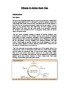
Preliminary experiment:
In this experiment I prepared and observed of the stages of mitosis in root tips. I did this by preparing a squash slide to view under a microscope. I understood the consequences of adding too much acetic orcein (stained chromosomes too deeply.) I heated the hydrochloric acid by warming over a flame; I developed this for my method, as it was not an accurate technique. The temperature couldn’t be controlled and therefore could disrupt my results so I plan on using a water bath at a set temperature and a thermometer to ensure the temperature was maintained.
Apparatus:
· Garlic clove – roots
· Microscope
· Nutrient solution
· Phosphate tablets - vary concentrations
· Acetic orcein
· Slides & coverslips
· Scalpel
· Watch glass
· Filter paper -mop excess liquids
· HCL (2.0 mol dm3)
· Thermometer
· Waterbath
· Test tubes
· Glass rod
· Volumetric flasks
· Beakers
Experiment Method:
1. Label 7 250cm volumetric flasks 0-5 & 10
2. Add one phosphate tablet to flask 1 and so on respectively. (flask 0 no tablets are added)
3. Fill flasks with distilled water, read at eye level and ensure that meniscus is resting on the line.
4. Label 7 500cm beakers 0-5 & 10.
5. Fill beakers with 200cm nutrient solution.
6. Add phosphate solution to each beaker respectively. Contents of flask 0 poured into beaker 0 e.t.c.
7. Cut 7 root tips 2cm long from garlic clove and place one into each beaker.
8. Leave for 48 hours.
9. To 7 test tubes add 10cm of hydrochloric acid and place in water bath at 60°C.
10. Transfer root tips to each test tube for 5 minutes.
11. Place root tip ‘0’ onto slides, cut apical tip 3mm long. Discard the rest.
12. Macerate root using glass rod and spread out as thinly as possible.
13. Add 3 drops of acetic-orcein allow to stain for 2 minutes.
14. Place coverslip on slide and blot firmly using filter paper; press gently to spread cells.
15. View under microscope.
16. Count how many cells in the 4 main stages of mitosis for each root.
17. Record results.
18. Repeat steps 11-17 for roots 1-5 and 10
19. Record results.
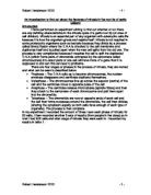
The recorded results shall go in a table like this:
(Using the results recorded in this table a graph can be plotted with x-axis “number of phosphate tablets used” and y-axis “ total number of cells in mitosis”, from this graph reliable and accurate conclusions can be drawn.)
Experiment Variables
Independent: different phosphate concentrations.
Dependent: number of cells undergoing mitosis.
Fixed/ confounding: (kept constant to ensure a fair test is carried out, and that the only variable having an effect on the rate of mitosis is the phosphate concentration.)
For all experiments:
1. Light – exposed to the same amount of light at all times (same time/same bench)
2. Temperature - carried out at room temperature.
3. Time- phosphate solution will be available to roots for 48 hours, ensuring fair results to be recorded for all tubes.
4. Nutrient solution – same amount will be used so results are not bias or disrupted.
5. Phosphate solution – amount used will be the same.
Experiment Risk Assessment