While some forms of meningitis are mild and resolve on their own, meningitis is a potentially serious condition owing to the proximity of the inflammation to the brain and spinal cord. The potential for serious neurological damage or even death necessitates prompt medical attention and evaluation. Infectious meningitis, the most common form, is typically treated with and requires close observation. Some forms of meningitis (such as those associated with , or infections) may be prevented with .
Headache is the most common symptom of meningitis (87 percent) followed by ("neck stiffness", 83 percent). The classic triad of diagnostic signs consists of nuchal rigidity, and altered mental status. All three features are present in only 44% of all cases of infectious meningitis. Other signs commonly associated with meningitis are (inability to tolerate bright light), (inability to tolerate loud noises), and (in small children) and (in 20-40% of cases). In infants (0-6 months), swelling of the (soft spot) may be present.
Investigations include (electrolytes, liver and kidney function, inflammatory markers and a ) and usually examination of the chest. The most important test in identifying or ruling out meningitis is analysis of the (fluid that envelops the brain and the spinal cord) through (LP).
The (CSF) sample is examined for (and which subtypes), , content and level. of the sample may demonstrate bacteria in bacterial meningitis, but absence of bacteria does not exclude bacterial meningitis; of the sample may still yield a causative organism. The Gram stain is positive in 60% of cases. Most cases of meningitis are caused by , such as , , , or , that spread into the blood and into the (CSF).
Staining mechanism
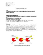
Gram-positive bacteria have a thick mesh-like cell wall made of (50-90% of cell wall), which stain purple and Gram-negative bacteria have a thinner layer (10% of cell wall), which stain pink. Gram-negative bacteria also have an additional outer membrane which contains , and is separated from the cell wall by the periplasmic space. There are four basic steps of the Gram stain, which include applying a primary stain () to a heat-fixed smear of a bacterial culture, followed by the addition of a (Gram's ), rapid decolorization with or , and counterstaining with or .
Gram staining protocol
- Make a slide of tissue or body fluid that is to be stained. Heat the slide for few seconds until it becomes hot to the touch so that bacteria are firmly mounted to the slide.
-
Add the primary stain and incubate 1 minute. This step colors all cells violet.
-
Add Gram's iodine, for 30 seconds. It is not a stain; it is a . It doesn't give color directly to the bacteria but it fixes the crystal violet to the bacterial cell wall. All cells remain violet.
- Wash with ethanol and acetone, the decolorizer. If the bacteria is Gram-positive it will retain the primary stain. If it is Gram-negative it will lose the primary stain and appear colorless.
-
Add the secondary stain, , and incubate 1 min, then wash with water for a maximum of 5 seconds. If the bacteria is Gram-positive then the cell will retain the primary stain, will not take the secondary stain, and will appear black-violet. If the bacteria is Gram-negative then the cell will lose the primary stain, take secondary stain, and will appear red-pink.
2-The ELISA technique
Enzyme-Linked ImmunoSorbent Assay or ELISA is a technique used mainly in to detect the presence of an or an in a sample. The ELISA has been used as a tool in medicine and , as well as a check in various industries. In simple terms, in ELISA an unknown amount of antigen is affixed to a surface, and then a specific antibody is washed over the surface so that it can bind to the antigen. This antibody is linked to an enzyme, and in the final step a substance is added that the enzyme can convert to some detectable signal. Thus in the case of fluorescence ELISA, when light is shone upon the sample, any antigen/antibody complexes will so that the amount of antigen in the sample can be measured.
Applications
Because the ELISA can be performed to evaluate either the presence of antigen or the presence of antibody in a sample, it is a useful tool both for determining antibody concentrations (such as with the or ) and also for detecting the presence of antigen.
In an ELISA test, a person's serum is diluted 400-fold and applied to a plate to which HIV antigens have been attached. If antibodies to HIV are present in the serum, they may bind to these HIV antigens. The plate is then washed to remove all other components of the serum.
A specially prepared "secondary antibody” an antibody that binds to human antibodies is then applied to the plate, followed by another wash. This secondary antibody is chemically linked in advance to an enzyme. Thus the plate will contain enzyme in proportion to the amount of secondary antibody bound to the plate. A substrate for the enzyme is applied, and catalysis by the enzyme leads to a change in color or fluorescence. ELISA results are reported as a number; the most controversial aspect of this test is determining the "cut-off" point between a positive and negative result.
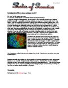
A 96-well being
used for ELISA.
3- Using the dilution plate technique
Much of the work of the clinical Microbiology laboratory is done to assist the clinician in the choice of drugs for the treatment of infections. It includes three main components:
- The identification of relevant pathogens in exudates which is body fluids (urine and faeces) collected from the patient.
- Sensitivity tests done to determine the degree of sensitivity or resistance of the pathogen isolated from the patient to an appropriate range of antimicrobial drugs which is antiseptics and antibiotics.
- Assays of the concentration of an administered drug in the blood or other body fluid of the patient required to control the schedule of dosage.
The clinician’s purpose in prescribing an antimicrobial drug is to produce at the site of infection a concentration of it high enough to kill or inhibit the growth of the responsible pathogen, without exerting a significant toxic effect on the patient’s tissues.
Sensitivity Tests
The results of sensitivity tests are generally reported in the form: ‘organism A isolated, sensitive (or resistant) to antibiotic B’ such a report implies that the organism is relevant to the patient’s clinical condition, that the minimum inhibitory concentration (MIC) of the antibiotic for it has been measured in some way, an that, if the organism is reported as ‘sensitive’, the MIC is less than half or a quarter the concentration of antibiotic likely to be found in the infected tissues of a patient given the usual schedule of doses, i.e. that the infection is treatable. A larger margin of safety may be required in some infections.
The MIC is measurable in three ways.
1. Tests with serial discontinuous concentrations. The organism is exposed to a series of fixed antibiotic concentrations in a separate culture in agar or broth. The different concentrations are produced by dilution, usually serially two-fold, of a solution prepared from antibiotic powder of accurately stated potency supplied for the purpose. They should not be prepared from therapeutic preparations, for the potency of these is not standardlised with sufficient precision.
The MIC is generally read as the smallest concentration of antibiotic in the series that prevents the development of visible growth of the test organism. The test can be extended to measure the minimum bacterial concentration (MBC) of the antibiotic by preparing subcultures from each test culture and nothing the smallest concentration of antibiotic from which no growth is obtained in the subculture.
-
Tests with break-point concentrations. This method is a modification of the foregoing one in which test organism, along with control strains of known MIC, is exposed to only two or three critical ‘break-point’ concentrations of the antibiotic in agar or both. These concentrations are chosen to reflect achievable antibiotic levels in the body fluids, usually blood and urine. If the organism is inhibited by the lower concentration it is reported as sensitive, if by the higher but not the lower, as of intermediate resistance and if not by the higher concentration as resistant.
-
Tests with diffusion gradients of concentration. By this method, the test organism is seeded uniformly over an agar surface and exposed to a continuous concentration gradient of antibiotic diffusing from a paper disk (disk diffusion test). A bacterium sensitive to the antibiotic is inhibited from growing in a circular zone around the paper disk; the lower the MIC of the antibiotic for the organism, the larger the diameter of the zone. A comparison of the zone size with that produced in a parallel test with a control strain gives a measure of the MIC.
Disk diffusion tests
The disk diffusion method is still the most widely used for the routine sensitivity testing of isolates from patients. It is satisfactory for organisms that grow well overnight, i.e. for most of the common pathogenic aerobic and facultatively anaerobic bacteria, rapidly growing anaerobes such as Clostridium perfringens and Bacteroides fragilis, and rapidly growing fungi such as Candida albicans.
It is unsuitable for slowly growing organisms, such as the tubercle bacillus, for the antibiotic would have become too diffused before growth had become visible.
Several forms of disk diffusion test have been advocated, which vary in their methods of standardisation, reading and control. Disk of a single, usually high content of antibiotic are placed on an inocullum of strictly standardized density on Muller-Hinton agar.