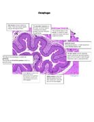
The next layer is submucosa. The submucosa is a thin layer of loose connective tissue that supports the mucosa; it also joins the mucosa to underlying muscle fibres. The submucosa has blood supply and nerves that is supplied to the mucosa.
Circular muscle is situated after the submucosa it is muscle that goes around the oesophagus. The Longitude muscular is situated after the circular muscle and goes along the oesophagus. Together they are called external muscle coat. The have a vital importers for peristalsis. Peristalsis is the movement of the alimentary canal that forces food continue on it journey. The external muscle coat contract in a wave like contraction above and under the bolus forcing food further down the alimentary canal.
The last layer is Serosa; it is a loose connective tissue.
Stomach: The oesophagus goes all the way down to the stomach. The real action starts here. The stomach is a sack like, oval shaped dilation of the alimentary canal. The Stomachs main function is to collect food, break the food down to a creamy liquid and release the food to the duodenum in small quantities. The stomach has the ability to expand when food arrives to the stomach, it also shrivels up when it is empty.
When the stomach is empty you can see with your eyes folds. The folds are named ruga; when the stomach is full these rugas irons out.
The stomach has a columnar epithelium, which has gastric pits. In the gastric pits are the gastric glands. The stomach is lined with mucus secreting cells that lines the stomach. Mucus is alkaline and vital for the stomach, as it needs protection from all the digestive juices so it does not self-digest. The gastric glands produce digestive juices and an enzyme called pepsin that breaks protein in to soluble solutions. The secretion of gastric juices are set of by smell or of sight of food by nerves impulses from the brain. The gastric juices are present as long as there is food. The epithelium and the muscularis mucosa are together commonly called gastric mucosa. The hydrochloric acid mixes with the rest of the gastric juice and makes a good environment for the pepsin to work and kills the bacteria that come with food. Beneath the gastric mucosa is muscularis mucosa and submucosa. The sub mucosa contains blood vessels and lymph vessels and nerve plexus. The blood vessels supply oxygen, remove carbon dioxide and absorb digested food. The next tissue is external muscle coat and serosa.
In the stomach the external muscle coat which is particularly important in the stomach, peristalsis action happens every 20 seconds to mix up the food. When food has broken down to liquid the pyloric sphincter releases the liquid little at the time to the duodenum.
Duodenum: duodenum is the first part of the small intestine. Most of digestion and absorption happens in the small intestine. The duodenum receives digestive enzymes from pancreas, pancreatic glands. (The enzymes are not active until they reach duodenum). The pancreatic juice contains sodium hydrogencarbonate, this is vital as the sodium hydrogencarbonate neutralizes the acid from the stomach, and enzymes don’t work well in acidy conditions. The pancreatic juice acts on all types of food. The duodenum also receives bile; it is produced in the liver and stored in the gall bladder. It is then pored in to the duodenum via ducts. The pancreatic juices enzymes are attacking all types of food. The bile assist in the process of dissolving fats. This gives the enzyme lipase a better surface to work on so the fat can be absorbed of the lining in the duodenum. The mucosa has adapted in the duodenum to maximise the intake of the absorption. The lining in the duodenum is a thin simple columnar epithelium covered by villi. They are finger or leaf like projections. They are 0.5mm in length, at the surface of each of the villus are fine hair called microvillus. The villus has specialised in absorbing food and, with the microvillus the surface is extended further. In the villus there are a fine network of capillaries transporting blood. Fluids can easily pass through the villus so they can absorb the liquid released from the stomach. The villuas also has mucus-secreting cells. In-between the villus are a depression in the surface, these depression is called crypt of lieberkun. They secrete an enzymes sucrose and maltase. In the bottom the crypt of lieberkuhn the Brunner’s gland are situated. The Brunners gland produce alkaline secretion in order to protect duodenum from acid from the stomach while providing a good environment for the enzyme to work, it also provide with protections for the intestinal walls. The hepatic portal vein is an amalgamation of veins that carry the blood from the intestine to the liver. After being filtered by the liver these products are released to the rest of the blood stream.
Ileum is the second part of the small intestine.
Ileums duty is to absorb all the nutrients from the duodenum that has been pushed along with peristalsis. The ileum is very effective in absorb the remained soluble products. The main reason for ileum is so good at it job is that ileum is longer that the duodenum and it has villus. The villus are smaller compared to the duodenum they have also smaller crypts. The ileum has paneth cells instead of Brunner’s gland. The paneth cells are important as they provide a defence against microbes. The paneth cells secrete antimicrobial molecules and that way protects the intestine from the microbes to enter the bloodstream. The villus has a complicated capillary system that transport absorbed products to the liver to be filtered. The villus also has lymph vessels or lacteal, these vessels are responsible for absorbing the fat that the bile and lipase broke down. The fats are transported into the lymphatic system and eventually emptied in the blood system. The lining in the epithelium is thinner so product can easily get absorbed.
Oesophagus adaptation is: the epithelium is stratified squamosis and non-katerised. That means that the oesophagus has a thicker layer epithelium. This is a vital adaptation as cells are constantly depleted by rough food or digestive enzymes, a layer of lost epithelium dose not have any effect on the oesophagus. The muscularis mucoae is thicker in the oesophagus than the rest of the oesophagus this to improve secretion of the mucosa and absorption. The oesophagus don’t have digestive enzymes glands, this is because digestion starts in the stomach, oesophagus main role is to transport food to the stomach.
Stomachs adaptation: The stomach most obvious adaptation is that the stomach swells out and act as storage until food has liquefied. The stomach has rugas that make it possible for the stomach to dilate when in work, or shrink when not in use. The lining is columnar epithelium and has gastric glands that produce digestive juices and acid, this make it possible for the stomach to do its job (process food for the next production line and easier to absorb). The adaptation of the epithelium is that the cells are thick and protective. The gastric pits make up the most of the epithelium. Peristalsis has adapted in the stomach to a more reoccurring action when food is in the stomach, movement happens every 20 sec. Pyloric sphincter is another important adaptations as it makes sure no solid pieces lets out in the duodenum.
Adaptations of the duodenum:
The duodenums main priority is to absorbed soluble products and have adapted in a way to fit the task there for it differentiate signifinically, the surface is villus to increase the surface aria, ducts from panaceas to transport enzymes to the duodenum, and bile ducts for assisting in dissolving fats. The duodenums epithelium is considerably thinner so the products can easily pass to the capillaries in the villi. The duodenum also produce sodium hydrogencarbonate neutralize acid from the stomach. The Brunner’s gland contains goblet cells that produce mucus to further protect the epithelium. The duodenum has a complicated blood system that transports the blood to the liver for filtering the blood before the blood enters the rest of the body.
Ileum specific adaptation: The villi in the ileum are narrower and the crypts are as deep as in the duodenum. But the ileum itself is much longer; it also has spiral rings throughout the whole ileum and effective villus. The villi are constantly shed in the lumen rapid cell division in the epithelium replaces the lost cells. The villi constantly renew them self and keep the epithelium health and effective. Each villus contains a lacteal that are a capillary of the lymphatic system that transport fat. The ileum also has paneth cells that protect the body from getting ill.
Conclusion: throughout the whole alimentary canal there are fundamentally similarities: The alimentary canal has epithelium, mucosa, muscularis mucosa, submucosa, circularmuscle, longitudinal muscle (together they are external muscular coat) and serosa. Other similarities are secreting mucas cells and peristalsis. The different part of the digestive system has adapted to make sure food is not damaging the alimentary canal and maximise the nutrion intake with many more important tasks. All this is done so we can function.
Source: GCSE biology third edition
www.medic8.com